Bile Acid Testing in Dogs: Why Timing and Feeding Instructions Matter
A guide to the canine bile acid challenge: why the 12-hour fast, test meal, and two-hour draw matter, how mistakes alter results, and how to read lab reference ranges.

What a bile acid test measures - and why it needs two timed samples
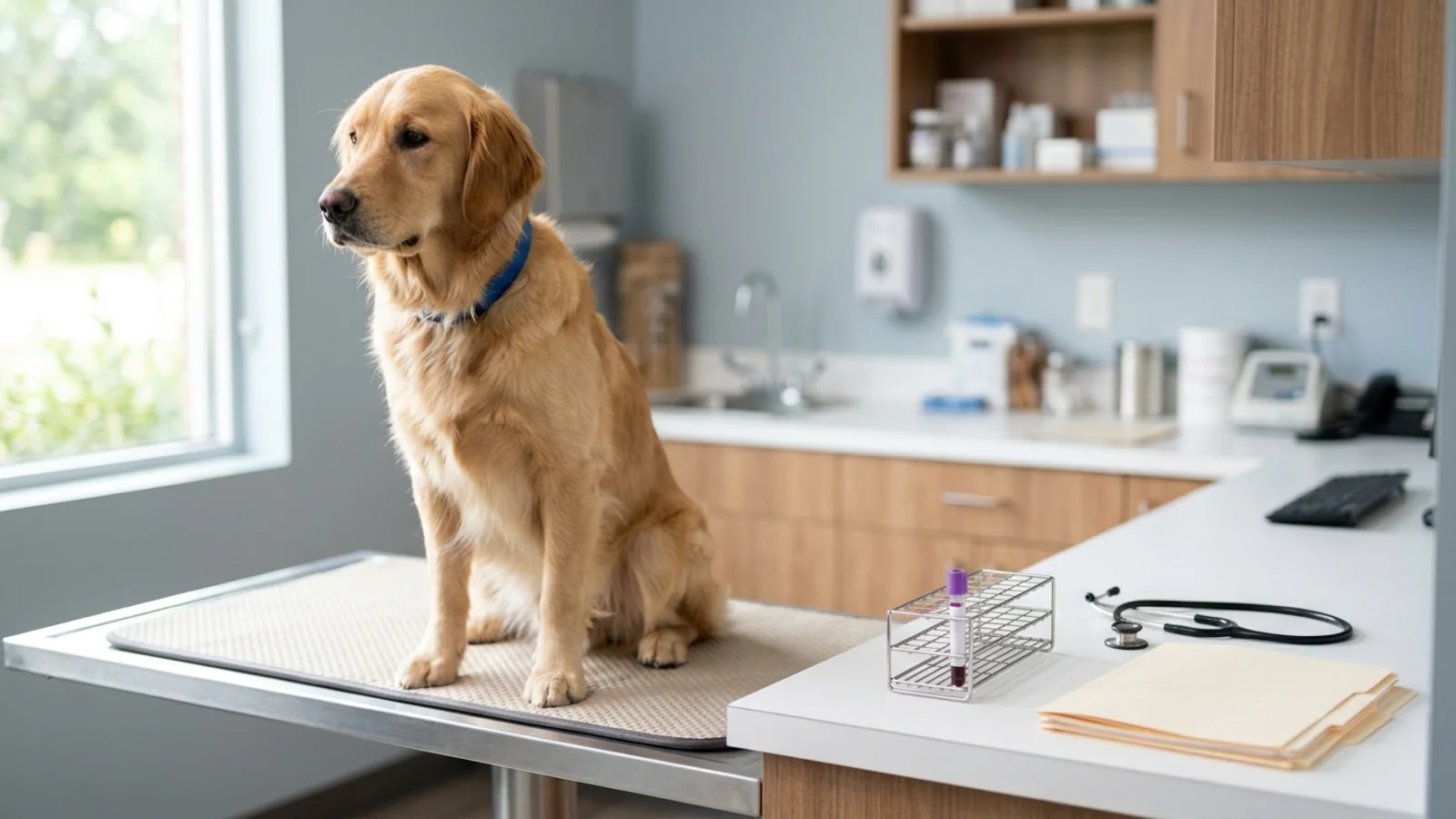
When a dog undergoes a canine serum bile acid test—frequently abbreviated as a total serum bile acid (TSBA) challenge—the veterinary team is not running a static chemistry check. Instead, they are conducting a dynamic, two-stage physiological challenge designed to evaluate functional liver mass and hepatic vascular circulation. The procedure requires two distinct blood draws: a resting preprandial sample collected after the fast the performing laboratory requests (commonly 8 to 12 hours; the Merck Veterinary Manual is the published outlier that does not require a fast), followed immediately by a measured test meal to trigger gallbladder contraction, and a final postprandial sample drawn precisely 120 minutes later. Each step in this timed sequence exists to challenge the liver's metabolic clearance capacity under controlled conditions.
To understand why two samples are necessary, pet owners and veterinary nurses must consider the mechanics of enterohepatic circulation. In a healthy canine, hepatocytes synthesize primary bile acids (chiefly cholic acid and chenodeoxycholic acid) from cholesterol. These acids are conjugated with amino acids—including taurine—and secreted through the bile canaliculi into hepatic ducts. Between meals, the sphincter of Oddi remains closed, directing bile into the gallbladder, where it is concentrated and safely stored. When food enters the stomach and passes into the duodenum, dietary fats and amino acids stimulate the release of cholecystokinin (CCK) from duodenal mucosal cells. CCK triggers rapid gallbladder contraction while relaxing the sphincter of Oddi, releasing a concentrated bolus of bile acids into the intestinal lumen to emulsify dietary lipids.
Once dietary fats are broken down and absorbed across the proximal jejunum, bile acids travel onward to the terminal ileum. There, specialized sodium-dependent active transport mechanisms reabsorb more than 90% of secreted bile acids into the portal vein. The portal venous system carries this high-concentration bile acid load directly back to the hepatic sinusoids. In a dog with normal liver microarchitecture and unimpeded portal blood flow, healthy hepatocytes extract most of those returning bile acids on the first pass through the sinusoids. Consequently, only a tiny fraction escapes into systemic peripheral blood. If the liver suffers from reduced functional mass (such as in cirrhosis or chronic hepatitis) or if portal blood bypasses hepatic sinusoids entirely via a vascular shunt, post-challenge bile acids spill over into peripheral circulation, resulting in dramatically elevated serum levels.
Pet owners often wonder why a standard blood panel is not sufficient. Routine serum biochemistries—such as those detailed in our comprehensive guide to canine and feline blood chemistry panels—measure circulating enzymes like alanine aminotransferase (ALT), aspartate aminotransferase (AST), alkaline phosphatase (ALP), and gamma-glutamyl transferase (GGT). While these biomarkers indicate active hepatocellular leakage or biliary ductal tension, they do not quantify how well the liver is actually functioning or whether blood is reaching the hepatic parenchyma. A dog with end-stage hepatic fibrosis or a severe congenital vascular anomaly can exhibit completely normal liver enzyme levels because there are few healthy hepatocytes left to leak enzymes. Conversely, dynamic bile acid testing directly interrogates functional clearance capacity.
In a 1991 Journal of the American Veterinary Medical Association study of 170 dogs evaluated for hepatobiliary disease (Center et al.), preprandial concentrations above 20 µmol/L and postprandial concentrations above 25 µmol/L each had 100% specificity for hepatobiliary disease in that study. The authors reported that a preprandial result above 20 µmol/L or a postprandial result above 25 µmol/L indicated histopathologic hepatobiliary abnormalities or a portosystemic vascular anastomosis. Overall efficacy of the two timed samples was nearly identical: 82.4% preprandial and 82.3% postprandial. Pairing them improved sensitivity for several groups, including cirrhosis, portosystemic vascular anomaly, and glucocorticoid hepatopathy. A 2021 multicenter retrospective study in the Journal of Veterinary Internal Medicine (Pena-Ramos et al.; 341 dogs with suspected liver disease and a liver biopsy) found postprandial bile acids more sensitive but less specific than resting values. At a cutoff of 10 µmol/L, specificity for all liver diseases combined was 49.3% for resting samples and 29.7% for postprandial samples. Cornell eClinPath, Cornell's Animal Health Diagnostic Center, and Gribbles still recommend the paired fasting-plus-two-hour panel because a single random sample can miss an abnormal value. The 1991 specificity figures came from that study's own control comparison; the 2021 series, using dogs with reactive hepatopathy as controls, shows why a single cutoff is not a universal verdict.
flowchart TD
    A["Fast requested by the laboratory, then preprandial draw"] --> B["Test meal specified by that same laboratory"]
    B --> C["Meal stimulates gallbladder contraction"]
    C --> D["Ileum reabsorbs most secreted bile acids"]
    D --> E["Portal blood returns the load to the liver"]
    E --> F{"Does the liver clear that load?"}
    F -->|"Efficient clearance"| G["Lower concentration in systemic blood"]
    F -->|"Reduced functional mass or a shunt"| H["More bile acids remain in systemic blood"]
    G --> I["Second draw two hours after the meal is finished"]
H --> IThe standard protocol: fast, draw, feed, wait two hours, draw again
A paired bile acid challenge is an exacting, timed diagnostic procedure. Executing the test requires strict coordination among the pet owner, the veterinary phlebotomist, and the clinical laboratory. Any deviation in fasting duration, food composition, or draw timing introduces preanalytical variance that can skew the final interpretation.
The timeline starts the evening before the appointment. IDEXX and Gribbles ask for about a 12-hour fast, and the Texas A&M GI Laboratory asks for 8 to 12 hours. VCA calls a 12-hour fast, including treats and chew toys, an important part of its protocol. Food, treats, chews, and flavored pill pockets are withheld because they can empty the gallbladder before the baseline sample. Fresh water stays available: Abbott Road Animal Hospital's client chart lists bile acids as a 12-hour fast with water allowed. Do not stop a prescribed medication in order to create that fast. If a dose cannot be given without food, call the clinic before the visit. The Merck Veterinary Manual does not require a fast; that disagreement is explained in the next section. Clinics usually book the first draw in the morning so the two-hour sample still fits in the day.
| Phase | Clock / Target | Patient State | Clinical Action | Diagnostic Purpose |
|---|---|---|---|---|
| 1. Pre-Test Fast | 8 to 12 hours prior | Fasted overnight; water allowed | Withhold food, treats, chews, and flavored pill pockets. Ask the clinic before giving or skipping any medication. | Clears postprandial circulation; pools bile in gallbladder; establishes clean baseline |
| 2. Baseline Draw (0 hr) | Hour 0:00 (Morning) | Calm, resting, fasted | Collect the serum volume the laboratory requests and label the tube 0 hr or preprandial. TVMDL asks for 0.5 mL serum, the Texas A&M GI Laboratory asks for 0.2 mL non-hemolytic non-lipemic serum, and Gribbles asks for at least 1 mL serum. | Measures resting serum bile acid concentration prior to stimulation |
| 3. Test Meal | Immediately post-draw | Eating under observation | Feed the meal the performing clinic or laboratory specified, and confirm the dog actually ate it. | Stimulates duodenal CCK release, initiating gallbladder contraction and bile release |
| 4. Timed Interval | 120-minute timer | Resting in quiet kennel or at home | Start timer when dog finishes eating; ensure no additional food is accessible | Allows transit, jejunal absorption, ileal transport, and hepatic first-pass uptake |
| 5. Postprandial Draw (2 hr) | Two hours after the meal is finished | Calm, post-digestive state | Collect second blood sample; label tube '2 hr / Postprandial' | Collects the two-hour sample the paired protocol is built around. It is the protocol time, not proof that every dog peaks at exactly 120 minutes. |
| 6. Specimen Handling | As soon as possible after clotting | Completed test | Separate serum from cells promptly and inspect for hemolysis or lipemia. | Cornell and eClinPath say lipemia falsely raises, and hemolysis falsely lowers, measured bile acids. Prompt separation limits both. |
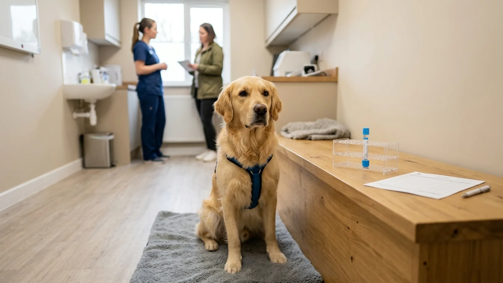
Veterinary diagnostic guidelines from the Cornell University Animal Health Diagnostic Center (AHDC) emphasize that paired bile acid testing is often best performed in an environment where the patient feels comfortable eating. In-hospital stress frequently causes dogs to refuse unfamiliar food or experience stress-induced gastrointestinal dysmotility. When a clinic permits home feeding, the pet owner brings the dog in for the fasting draw, returns home to administer the designated breakfast at routine feeding time, and returns to the hospital exactly two hours after meal completion for the second phlebotomy. Regardless of setting, recording the precise minute the dog finishes eating is paramount.
Why the fast matters - and what a sneaked treat does to the baseline number
The primary physiological objective of the 8- to 12-hour fast is to achieve a state of hepatobiliary quiescence. In the interdigestive state, biliary synthesis continues at a basal rate while the gallbladder stores and concentrates hepatic secretions. In healthy dogs, circulating resting bile acids remain low—typically well below 15 µmol/L on general university diagnostic panels and 6.0 µmol/L or less on Texas A&M GI Laboratory intervals.
If a patient consumes food during the fasting window—whether a handful of morning kibble, a dental chew stick, or a pill pocket—the enterohepatic cycle initiates prematurely. Gastric distension and peptide presence trigger duodenal CCK release, prompting the gallbladder to discharge its contents into the bowel. When the veterinary phlebotomist collects what is assumed to be a 'fasting' sample, the blood actually reflects an unstandardized, partial postprandial state. The resting number reads falsely elevated, obscuring whether an elevated 2-hour sample represents genuine hepatic insufficiency or merely the continuation of an uncoordinated meal cycle.
Abbott Road Animal Hospital's fasting chart is one clinic's instruction, and it matches the fast-plus-water pattern used by the laboratories above: bile acids are listed as a 12-hour fast with water allowed, and a morning drop-off. The same sheet says that, for blood tests in general, a difficult medication may be given with a small amount of food. That exception works against this test, because any food can start gallbladder contraction before the baseline draw. If a dose cannot be given without food, call the clinic rather than improvising a snack. The chart does not say that withholding water improves a bile-acid result.
Feeding instructions: why labs disagree about the test meal
Perhaps no aspect of canine bile acid testing generates more client confusion—and veterinary protocol debate—than the composition and volume of the stimulation meal. Different reference laboratories and diagnostic textbooks publish contradictory feeding recommendations, leaving practitioners and pet owners to navigate competing instructions.
The physiological goal of the test meal is straightforward: deliver sufficient dietary fat and protein to the duodenal lumen to stimulate robust CCK secretion and comprehensive gallbladder contraction. However, laboratory opinions diverge sharply on how much dietary fat is necessary to achieve this response without creating specimen interference:
| Institution / Source | Recommended Test Meal | Portion Size Guidance | Underlying Rationale | Reported Technical Caution |
|---|---|---|---|---|
| IDEXX Laboratories (Catalyst) | High-fat commercial or veterinary diet | Minimum 2 tsp for dogs <10 lb; 2 tbsp for larger dogs | IDEXX states that a high-fat meal is used to stimulate gallbladder contraction. It also gives a minimum amount, and an option to mix a little corn oil into a restricted-protein food if protein is a concern. | IDEXX does not publish a lipemia warning on this protocol page. Cornell and Gribbles warn that too large or too fatty a meal can lipemize the sample. |
| Gribbles Veterinary Pathology | Standard canned maintenance diet | 2 to 4 tablespoons total volume | Gribbles says a high-fat diet is not necessary. A canned maintenance diet is the stimulus they specify. | Gribbles says a high-fat diet can contribute to unwanted lipemia if given in excess, and that lipemia interferes with measurement. |
| Cornell University (AHDC) | Patient's regular commercial diet | Routine meal quantity and composition | Cornell says to feed the regular meal, type and amount, at the usual meal time, so the dog is more likely to eat and to have normal gastrointestinal motility. Home feeding is often best for that reason. | Cornell's published caution about a large meal is lipemia, which falsely increases the result, not a stated rule that extra volume always delays gastric transit past two hours. |
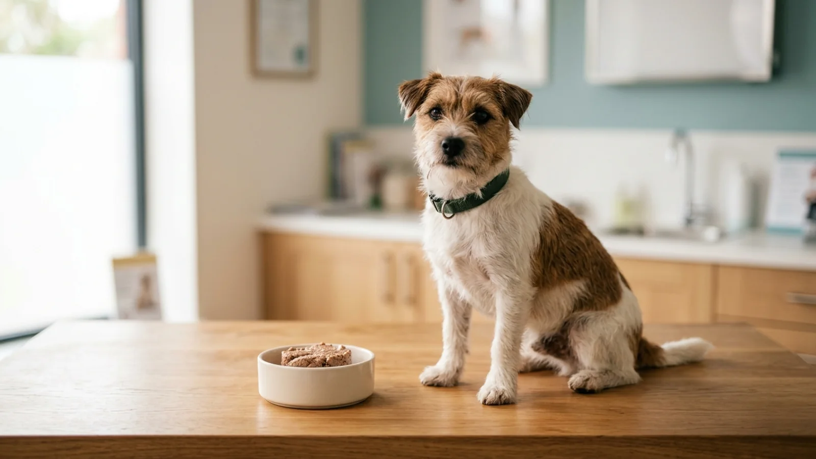
| VCA Animal Hospitals | Palatable canned loaf or stew | A small tasty meal of canned food, eaten promptly | VCA describes a small canned meal after the resting sample, then a second sample exactly two hours after the meal is finished. | VCA says failing to fast, feeding too large a meal, feeding dry kibble rather than canned food, or drawing blood at the wrong time can affect validity. It does not publish a gastric-emptying time for kibble. |
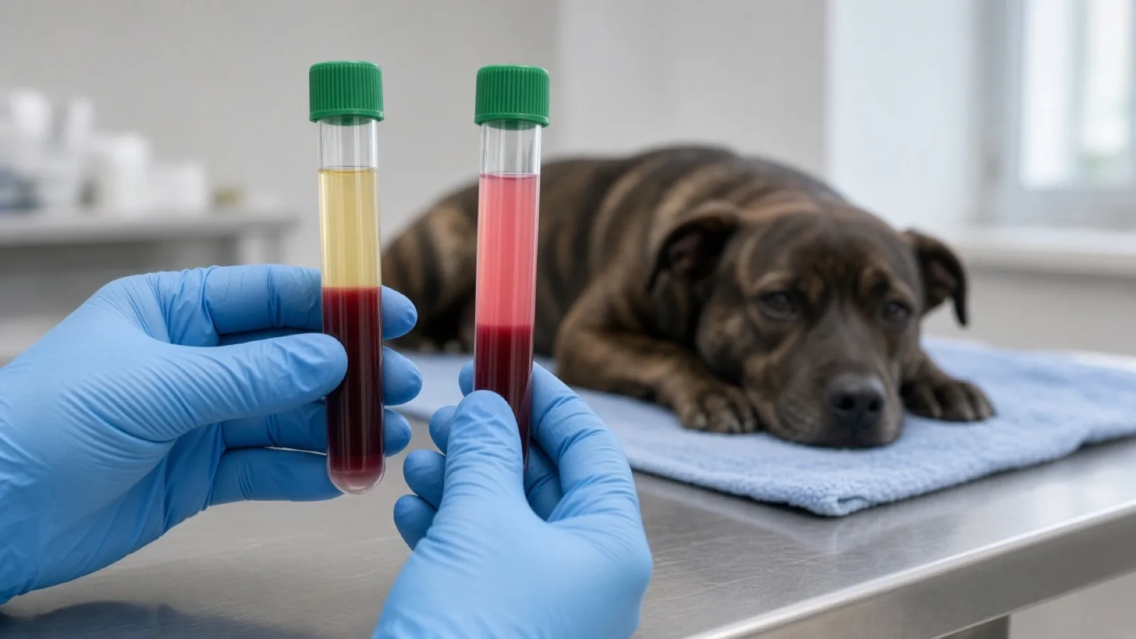
The main analytical problem with a meal that is too large or too fatty is preanalytical lipemia. Cornell's Animal Health Diagnostic Center and eClinPath state that lipemia falsely increases measured bile acids. eClinPath attributes that effect to optical interference with older reagents and cites Solter et al., 1992. The same page notes a manufacturer application sheet claiming that triglyceride up to 750 mg/dL, and hemoglobin up to 500 mg/dL, should not substantially interfere, and eClinPath says that claim has not been formally tested in animal samples. Cornell's practical instruction is still to avoid an overly large meal and to separate serum from cells as soon as possible. A milky postprandial sample is a reason to question the number, not a diagnosis of liver disease.
VCA lists feeding dry kibble rather than canned food as a protocol error that can affect validity. eClinPath separately lists delayed gastric emptying among the reasons a bile-acid concentration can be falsely low, because the meal may not challenge the liver during the two-hour window. Those cautions belong together, but they are not proof that every kibble meal is still in the stomach at 120 minutes. Feed the food the laboratory named. Tell the clinic the brand, the texture, the amount, and whether any of it was refused or vomited.
Timing and handling errors: which mistakes push the result up or down
Interpreting a canine bile acid profile requires clinical confidence that the numerical values reflect genuine physiology rather than laboratory artifacts. Protocol mistakes, specimen handling errors, and patient variables alter bile acid concentrations in predictable, known directions. Understanding these error vectors allows veterinary teams and pet owners to avoid confounding mistakes.
| Protocol Deviation / Variable | Biological Mechanism | Effect on Fasting (0 hr) | Effect on Postprandial (2 hr) | Net Diagnostic Risk |
|---|---|---|---|---|
| Broken Fast / Sneaked Treats | Premature CCK release and partial gallbladder emptying before baseline draw | Falsely Increased (High Baseline) | Unpredictable / Blunted Ratio | False-positive fasting elevation; masks true dynamic response to meal |
| Excessively Fatty / Large Meal | Lipemia from a large or very fatty test meal causes optical interference. eClinPath and Cornell say this falsely increases the measured concentration. | No Direct Effect | Falsely Increased (Lipemia Artifact) | A lipemic increase can look like hepatobiliary disease. Ask the laboratory whether the sample was flagged. |
| Traumatic Phlebotomy / Hemolysis | Lysis of RBCs releases free hemoglobin, absorbing spectrophotometric light | Falsely Decreased | Falsely Decreased | False-negative result; can mask significant shunts or hepatic failure |
| Second draw well before 2 hours | VCA says the wrong draw time can affect validity. The cited pages do not publish a less-than-90-minute false-negative cutoff. | No Direct Effect | May no longer match the two-hour interval the laboratory uses. Direction is not a published minute rule. | The sample is not the two-hour sample the protocol describes. Tell the clinic the actual clock time. |
| Second draw several hours late | eClinPath says rapid gastrointestinal transit can lower concentrations. The cited pages do not define a greater-than-3-hour cutoff. | No Direct Effect | May be lower than the two-hour sample would have been. Not a published clock rule. | A late sample can understate the challenge. Record the actual interval. |
| Meal Refusal / Vomiting | Absence of duodenal fat/protein stimulus; no gallbladder contraction occurs | No Direct Effect | Falsely Decreased (No Stimulus) | False-negative challenge; failure of dynamic test execution |
| Delayed Gastric Emptying | Food trapped in stomach; bile release delayed past the 120-minute draw | No Direct Effect | Falsely Decreased (Delayed Peak) | False-negative test; patient may show inverted fasting > postprandial |
| Puppy <16 Weeks of Age | eClinPath says concentrations may be falsely lower before 16 weeks of age in breeds predisposed to congenital portosystemic shunts. The note is attributed to Dr. Center by personal communication, not a consensus statement. | May be falsely low | May be falsely low | A low result in a young puppy of a predisposed breed is hard to trust. eClinPath says test after 16 weeks. The clinic decides timing. |
| Ursodiol (UDCA) Therapy | Merck states that ursodeoxycholic acid is measured by the bile acid assay and can increase the measured total. | Can be increased | Can be increased | The number may reflect the drug. Any pause is the prescriber's decision, not an owner instruction. |
| Clinical Icterus / Cholestasis | Cornell AHDC: icterus from cholestasis, or biochemical cholestasis with high total and direct bilirubin and bilirubinuria, already shows the pathway is not clearing bile normally. | Not useful for function or shunting | Not useful for function or shunting | Cornell says the test adds no information about function or vascular abnormalities in that setting. |
Specimen hemolysis represents a major source of analytical error in canine diagnostics. As discussed in our analysis of blood sample hemolysis in dogs, traumatic phlebotomy, excessive syringe suction, or forcing blood through a small needle can lyse erythrocytes. eClinPath and Cornell's Animal Health Diagnostic Center state that hemolysis falsely decreases measured bile acids through optical interference with older methods. Gribbles says to limit that interference by fasting, using a large-gauge needle, preferring jugular collection, and separating serum after clotting. Gribbles does not publish a 20- or 21-gauge requirement.
Cornell's Animal Health Diagnostic Center says bile acids should not be run in a dog with icterus from cholestasis, or with biochemical cholestasis (high total and direct bilirubin plus bilirubinuria). In that setting the test adds no information about hepatic function or a portosystemic shunt. A canine gallbladder mucocele is one disease that can reach obstructive cholestasis. Whether to test, image, or operate is the clinician's decision. Jaundice from red-cell destruction is a different problem and is not the cholestasis exclusion Cornell describes.
Reading the two numbers: why the same value is 'high' at one lab and normal at another
The same integer changes meaning across laboratories. A postprandial result of 26 µmol/L sits in the Texas A&M GI Laboratory's mildly increased postprandial band (13.6 to 29.9 µmol/L, described as usually not clinically significant), above the Merck Veterinary Manual's abnormal threshold of 25 µmol/L, inside Gribbles' equivocal band of 15 to 31 µmol/L, and in Cornell's suggestive range above 25 to 30 µmol/L. Read the pair against the laboratory that reported it.
| Diagnostic Institution | Preprandial guidance | Postprandial guidance | Intermediate zone, as published | Result the source treats as abnormal |
|---|---|---|---|---|
| Texas A&M University (GI Lab) | Reference interval ≤ 6.0 µmol/L. From 6.1 to 14.9 µmol/L is mildly increased and usually not clinically significant. A result inside the interval does not rule out liver disease. | Reference interval ≤ 13.5 µmol/L. From 13.6 to 29.9 µmol/L is mildly increased and usually not clinically significant. A result inside the interval does not rule out liver disease. | The 13.6 to 29.9 µmol/L band is the postprandial "usually not clinically significant" zone. It is not the fasting cutoff. | Preprandial ≥ 15 µmol/L, or postprandial ≥ 30 µmol/L, suggests significant hepatobiliary dysfunction or abnormal perfusion. Texas A&M says this suggests the problem and does not conclusively prove it. |
| Cornell University (AHDC) | No separate fasting reference interval of ≤ 15 µmol/L is published as a cutoff. eClinPath says fasting samples are generally under 15 µmol/L. Cornell applies the same guidelines to fasting, postprandial, and random samples. | Same interpretive guidelines as the fasting and random samples. There is no distinct postprandial reference interval of ≤ 15 to 25 µmol/L. | 15 to 25 µmol/L is equivocal in Cornell's summary of Dr. Center's biopsy studies: dogs in that range may or may not have hepatic pathology. | Above 25 to 30 µmol/L is suggestive of hepatobiliary disease. Cornell's summary of those biopsy studies says dogs under 25 µmol/L did not show hepatic pathology and dogs over 25 usually did. The 2021 Journal of Veterinary Internal Medicine series found lower specificity and found liver disease with resting values under 10 µmol/L, so Cornell's zones are Cornell's guidance, not a guarantee. |
| Gribbles Veterinary Pathology | The Gribbles protocol page does not publish a canine fasting reference interval of ≤ 15 µmol/L. It does say hepatobiliary dysfunction is unlikely when both results are under 31 µmol/L, especially under 15. | Postprandial values are the ones Gribbles scores against 31 µmol/L. A postprandial reference interval of ≤ 15 µmol/L is not what the page states. | 15 to 31 µmol/L is equivocal: the dog may or may not have liver dysfunction. | Postprandial above 31 µmol/L is suggestive of decreased functional mass, cholestasis, or portovascular shunting. If both results are under 31 µmol/L, especially under 15, Gribbles says dysfunction is unlikely. |
| Merck Veterinary Manual | Fasting ranges should not be applied. Fasting is not required for the procedure. A result above 25 µmol/L is abnormal before or after the meal. | Above 25 µmol/L is abnormal after the meal as well. Merck does not publish a separate normal postprandial ceiling of ≤ 25 as a fasting-style reference interval. | No equivocal zone is defined in this chapter. | Above 25 µmol/L in dogs, on either the pre-meal or the two-hour sample. Cats are a different threshold in the same chapter (above 20 µmol/L) and are outside this dog-focused comparison. |
eClinPath says to use the higher of the two concentrations, rather than assuming the postprandial result will always be higher. A fasting result of 42 µmol/L with a postprandial result of 28 µmol/L is an illustration, not a patient: at the Texas A&M GI Laboratory the fasting value would already sit at or above the 15 µmol/L preprandial flag, while 28 µmol/L would still be in the mildly increased postprandial band. Which sentence the report prints depends on which laboratory ran the assay.
When the fasting number is higher than the two-hour number
One of the most common sources of panic for pet owners—and confusion for early-career clinicians—occurs when the fasting bile acid value exceeds the 2-hour postprandial value. For example, an owner might receive a report showing a preprandial bile acid of 11.2 µmol/L and a postprandial bile acid of 4.8 µmol/L. Intuition suggests that after eating, bile acids should inevitably climb.
Merck reports that about 15% to 20% of dogs, and 5% to 10% of cats, have a higher total serum bile acid concentration before the meal than after. Cornell's Animal Health Diagnostic Center says this pattern occurs in up to 20% of dogs, from a recent meal, gallbladder contraction during the fast, or delayed gastric emptying. Gribbles says the fasting result is higher up to 20% of the time, and adds insufficient gallbladder contraction plus variation in intestinal transit or absorption. The cat figure is Merck's, stated here only so a mixed-species textbook sentence is not mistaken for a canine rate.
A recent meal or gallbladder contraction during the fast: Cornell and Gribbles both list a recent meal and spontaneous gallbladder contraction during the fast. The morning sample is then not a resting baseline.
Delayed gastric emptying: Cornell, Gribbles, and eClinPath list this. If the meal is still in the stomach at the two-hour draw, the intestine may not have been challenged yet, and the postprandial number can be lower than the fasting number.
Transit, absorption, and a fast that was too long: eClinPath says prolonged fasting, intestinal malabsorption, and rapid gastrointestinal transit can lower concentrations and reduce sensitivity. Gribbles includes variation in intestinal transit and absorption. None of these pages defines a 45- to 60-minute peak.
Cornell says that when the fasting value is the higher one and both results are under 25 µmol/L, especially under 15 µmol/L, hepatobiliary disease is unlikely. Gribbles uses a wider pair: both under 31 µmol/L, especially under 15. The Texas A&M GI Laboratory states the other limit directly: a result inside the reference interval does not rule out liver disease. An inverted pair is not, by itself, a disease. It is also not proof that the liver is normal or that further testing is unnecessary. The clinician reads both numbers on that laboratory's own scale.
Special Breed Considerations: The Maltese Update
Gribbles previously questioned bile-acid testing in Maltese dogs because a 1995 Australian study (Tisdall et al.) found high enzymatic results in many Maltese, with liver biopsies in only 11 of 200 dogs, and raised the possibility of an interfering substance. Gribbles now cites research presented by Dr. Sharon Center at the 2012 ACVIM Forum. Among 136 Maltese liver biopsies in that report, 22% had a classic congenital portosystemic vascular anomaly and about 60% had microvascular dysplasia without that anomaly. Every Maltese in that series who had portal hypoperfusion also had high serum bile acids, and Gribbles notes that a high ALT together with high bile acids is what usually prompted the biopsy. Those percentages describe a biopsied group, not the prevalence in all Maltese dogs. Gribbles says it now endorses the challenge test in Maltese dogs as in other breeds. This is Gribbles' summary of conference proceedings, not a Cornell Animal Health Diagnostic Center guideline and not a population survey.
What the test can and cannot tell you
A paired bile acid test is a powerful screening tool, but it is not an all-inclusive diagnostic verdict. Clarifying the test's scope and limitations prevents misdirected diagnostic journeys and unrealistic expectations.
What the test successfully detects:
Reduced Functional Hepatic Mass: Cornell and Gribbles list decreased functional hepatic mass, cholestasis, and portovascular shunting as separate reasons a result rises. In the 2021 Journal of Veterinary Internal Medicine biopsy series, the highest median resting concentration was in cirrhosis. A shunt can raise bile acids by diverting portal blood even when the dog is not in end-stage synthetic failure. These cited pages do not give a percent of liver tissue that must be lost before the test becomes abnormal.
Portosystemic Vascular Shunting: Abnormal vascular channels let portal blood bypass hepatic sinusoids. Cornell and Gribbles say most animals with congenital or acquired shunts have markedly increased postprandial bile acids. In the 2021 Journal of Veterinary Internal Medicine series, the median postprandial concentration in congenital circulatory anomalies was 126 µmol/L, with a range from 0 to 726 µmol/L, so a shunt can also be present with a normal result. Surgery and medical-management decisions are covered in the guide to portosystemic shunts in dogs.
Hepatic Microvascular Dysplasia (MVD): Microscopic intrahepatic portal hypoperfusion that impairs sinusoid delivery without a gross macroscopic shunt.
Subclinical Biliary Cholestasis: Early impairment of bile acid secretion before systemic hyperbilirubinemia develops.
What the test CANNOT do:
It cannot provide an etiologic diagnosis: An elevated result shows a clearance or perfusion problem. It does not separate a congenital shunt, microvascular dysplasia, and other hepatobiliary diseases. An example number such as 85 µmol/L is not a special diagnostic tier.
It cannot assess disease reversibility: A severely elevated number does not indicate whether the underlying condition is treatable, progressive, or reversible.
A normal result does not 100% exclude liver pathology: VCA and the Texas A&M GI Laboratory both say a normal or in-interval result does not fully exclude liver disease. Focal or mild disease can leave enough functioning tissue to clear the challenge. The 2021 series found dogs in every liver-disease category with resting values under 10 µmol/L and over 90 µmol/L.
eClinPath states that a high bile acid concentration is not specific for liver dysfunction or abnormal portal flow. In one study of 15 dogs with hyperadrenocorticism, fasting concentrations reached 118 µmol/L; 53% had high fasting values and 40% had high postprandial values (Tinted et al., 2023, as summarized by eClinPath). Concentrations did not fall significantly after trilostane in that summary, and a subset of healthy dogs in the same study had abnormally high values. eClinPath describes the increase as glucocorticoid hepatopathy from glycogen accumulation; the study authors attributed it to cholestasis even though bilirubin did not rise. eClinPath also gives dental disease in dogs as an example of a high result without other clinical or laboratory evidence of liver disease, attributed to Dr. Center by personal communication. A high number is a screening signal. It is not a diagnosis of primary liver failure.
When a result is abnormal, the veterinarian chooses the next step. The items below are examples grounded in the sources above, not a sequence this page is prescribing.
Fasting Abdominal Ultrasound: Evaluates hepatic size, parenchymal echogenicity, biliary architecture, and portal vessel flow. Preparation requires a specific imaging fast, detailed in our guide to dog ultrasound fasting protocols.
Coagulation Profiling: Prothrombin time (PT) and activated partial thromboplastin time (aPTT) evaluate hepatic synthesis of clotting factors before invasive procedures.
Other clinician-chosen tests: Some shunt workups include protein C activity, coagulation testing before a liver biopsy, or vascular imaging. This article does not quote a protein C percentage. The shunt guide covers that decision, and the veterinarian chooses the next test.
Advanced Vascular Imaging: Computed tomography angiography (CTA) or Doppler ultrasound to definitively map abnormal vascular anatomy.
Hepatic Biopsy and Histopathology: Center and colleagues treated abnormal bile acids as a reason to consider hepatic biopsy, not as a substitute for a tissue diagnosis. Whether to biopsy is the clinician's decision.
TVMDL publishes a fee of 21.00 USD per sample for automated bile acid measurement and states that the listed fee is per sample. A paired test uses two samples. Two listed fees come to 42.00 USD only if both samples are billed at that catalog rate. That arithmetic is not a package price, and it is not a clinic invoice. Hospital charges for the visit, the blood draws, the meal, and interpretation are separate and are not estimated here.
Owner checklist: what to write down and hand to the clinic
Because preanalytical variables directly govern the accuracy of bile acid testing, detailed owner record-keeping provides immense clinical value to the veterinary medical team. Arriving at the appointment with an accurate written log allows the attending clinician to interpret borderline numbers with confidence.
| Clinical Parameter | Recommended Log Entry | Diagnostic Value to Clinician |
|---|---|---|
| Fast Initiation Time | Exact time evening food was removed (e.g., 8:00 PM) | Confirms whether the patient achieved the required 8 to 12 hour fasting baseline |
| Water Access | Confirm water remained accessible overnight | Verifies patient hydration state and rules out hemoconcentration artifacts |
| Accidental Food / Treat Ingestion | Record any sneaked crumbs, chew toys, or treats, with exact time | Identifies premature gallbladder contraction if fasting baseline returns elevated |
| Current Medication Regimen | List all oral medications, especially Ursodiol, steroids, or liver supplements | Detects assay cross-reactivity (Ursodiol) or endocrine-induced elevations |
| Stimulation Meal Administration | Diet brand, food texture (canned vs kibble), and exact volume consumed | Confirms adequate protein/fat challenge and assesses lipemia risk |
| Meal Finish Timestamp | Precise minute the dog consumed the last bite (e.g., 8:42 AM) | Establishes the exact 120-minute countdown for the postprandial blood draw |
| Gastrointestinal Observations | Note any lip-smacking, nausea, food refusal, regurgitation, or vomiting | Identifies incomplete gallbladder stimulation or delayed gastric emptying |
| Tube Label Verification | Verify clinic labeled tubes '0 hr / Preprandial' and '2 hr / Postprandial' | Eliminates lab submission mix-ups where sample order is accidentally reversed |
When the test is completed, remember that laboratory values are one component of a holistic clinical assessment. While learning that a bile acid concentration is elevated can feel daunting, the test provides your veterinary team with the physiological evidence needed to protect your dog's health. Work closely with your veterinarian to interpret these numbers in the context of your dog's physical exam, history, and imaging findings.
Sources
1. Cornell University College of Veterinary Medicine (eClinPath). Bile acids - Clinical Pathology Laboratory Reference. Reference on enterohepatic circulation, using the higher of the two results, lipemia and hemolysis direction, non-hepatic increases, and the under-16-weeks note attributed to Dr. Center by personal communication.
2. Cornell University Animal Health Diagnostic Center (AHDC). Serum Bile Acids Results: Guidelines for Interpretation. Diagnostic laboratory guidance on the paired protocol, feeding the regular meal (often at home), lipemia and hemolysis direction, the 15–25 µmol/L equivocal zone, suggestive values above 25–30 µmol/L, and not testing patients with cholestasis.
3. Merck Veterinary Manual. Hepatic Function Tests in Small Animals. Textbook reference covering total serum bile acid physiology, the 15–20% incidence of fasting values exceeding postprandial values, ursodiol assay cross-reactivity, and screening protocols for predisposed breeds.
4. VCA Animal Hospitals. Bile Acid Test Client Education. Client-facing diagnostic summary detailing the 12-hour fast, test meal consistency, canned food versus dry kibble considerations, and next steps following abnormal results.
5. Gribbles Veterinary Pathology. Bile Acids Testing in Dogs and Cats. Commercial veterinary laboratory protocol covering the 12-hour fast, 2–4 tablespoon canned maintenance diet recommendation, preanalytical lipemia warnings, 0 hr / 2 hr tube labeling, and Maltese breed diagnostic validity.
6. Texas A&M University Gastrointestinal Laboratory. Serum Bile Acids Assay Guidelines. Canine reference intervals are preprandial ≤ 6.0 µmol/L and postprandial ≤ 13.5 µmol/L. Preprandial ≥ 15 µmol/L, and postprandial ≥ 30 µmol/L, suggest significant dysfunction or abnormal perfusion but do not prove it. Postprandial 13.6 to 29.9 µmol/L is mildly increased and usually not clinically significant. A result inside the interval does not rule out liver disease.
7. Texas A&M Veterinary Medical Diagnostic Laboratory (TVMDL). Bile Acid Test Catalog and Submission Specifications. Official diagnostic laboratory listing detailing specimen volume requirements (0.5 mL serum), sample labeling specifications, and public per-sample fee schedule (21.00 USD per sample).
8. IDEXX Laboratories. Catalyst Bile Acids Product and Protocol Specifications. Point-of-care analyzer protocol detailing the manufacturer's recommended high-fat stimulation meal guidelines, test workflow, and measurement range (1.0–180.0 µmol/L).
9. Center SA, ManWarren T, Slater MR, Wilentz E. Evaluation of twelve-hour preprandial and two-hour postprandial serum bile acids concentrations for diagnosis of hepatobiliary disease in dogs. Journal of the American Veterinary Medical Association, 1991;199(2):217-226. In that study of 170 dogs, preprandial values above 20 µmol/L and postprandial values above 25 µmol/L each had 100% specificity for hepatobiliary disease.
10. Pena-Ramos J, et al. Resting and postprandial serum bile acid concentrations in dogs with liver disease. Journal of Veterinary Internal Medicine, 2021;35(3):1333-1341. Retrospective study of 341 dogs with suspected liver disease and a liver biopsy. Postprandial samples were more sensitive and less specific than resting samples.
11. Abbott Road Animal Hospital. Veterinary Fasting Instructions. General veterinary practice client guideline demonstrating routine clinical protocol for 12-hour bile acid fasting with unrestricted water access.