A Hemolyzed Blood Sample: Why the Vet May Need Another Draw
When a dog's blood sample hemolyzes, ruptured red cells can distort specific results. See which values stop being trustworthy, why the vet may ask for another draw, and what the flag does not prove.

What Hemolyzed Actually Means on a Dog's Lab Report
Receiving a follow-up phone call from your veterinary clinic reporting that your dog's blood sample was hemolyzed—and that the veterinary team needs to bring your dog back for another blood draw—frequently prompts an understandable spike of anxiety. For many pet owners, medical terminology heard over the phone sounds inherently ominous. The immediate fear is that the dog has developed an acute blood cancer, a life-threatening bone marrow disorder, or a sudden collapse in organ function.
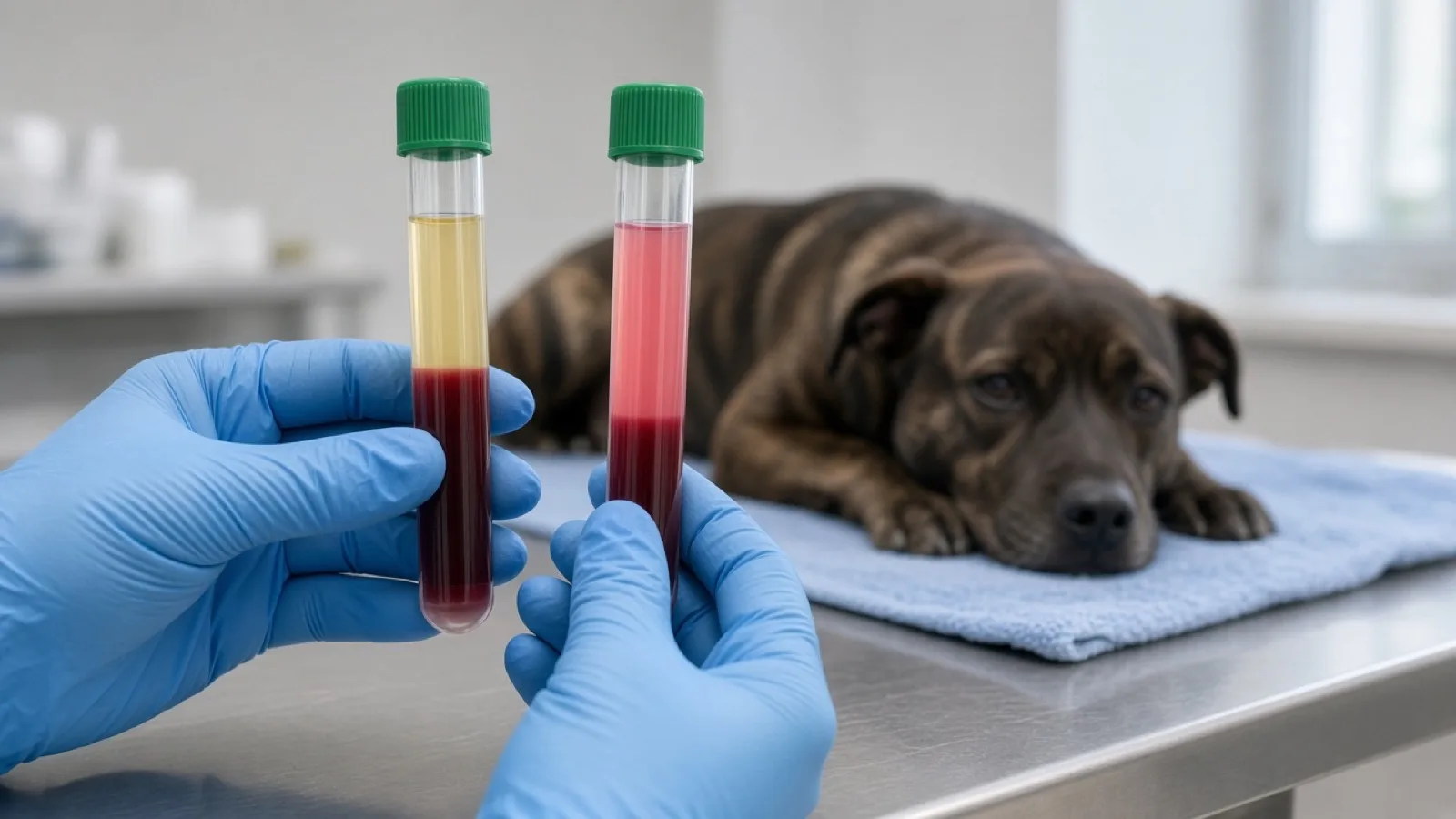
That fear is usually misplaced. A hemolyzed sample is a specimen quality problem, not a medical diagnosis. In clinical pathology, hemolysis refers to the physical rupture or lysis of erythrocytes (red blood cells), resulting in the leakage of their internal contents into the surrounding fluid matrix—the serum or plasma. Under normal circumstances, after whole blood is spun down in a laboratory centrifuge, the dense red blood cells form a solid packed pellet at the base of the tube, leaving a clear, pale straw-yellow fluid on top. When a sample hemolyzes, however, hemoglobin and intracellular constituents escape into that fluid, staining it distinct shades ranging from delicate light pink to deep port-wine cranberry red.
Cornell University's eClinpath states that hemolysis is the most common interference in clinical pathology testing. It is mostly from collection or handling, though it can also come from an intravascular hemolytic anemia. The page cites Whipple et al. 2020 for that frequency. Purdue's animal-disease laboratory describes hemolysis as red cells breaking because a sample was mishandled during collection or transport, and it does not estimate how often that happens. Most of the time, the pink serum is an in vitro artifact: damage inside the needle, syringe, tube, centrifuge, or shipping pack after the blood has left the vein.
If your dog is acting the way they did when they left the clinic, the phone call is about the sample. Free hemoglobin is deeply colored and chemically active. In the serum it changes how optical chemistry tests read, it can inhibit some reactions, and it releases substances that were inside the red cells. The numbers on that sample may no longer match what is circulating in your dog. Whether every test must be redrawn, or whether some values can still be used, is a test-by-test decision for the treating veterinarian.
How a Sample Hemolyzes: From Needle to Mailer
To understand why a blood sample hemolyzes, one must appreciate the delicate biophysical structure of the canine red blood cell. Erythrocytes are flexible biconcave discs bounded by a fragile lipid bilayer membrane. While engineered to deform smoothly through microscopic capillary beds under physiological blood pressure, they are remarkably vulnerable to sudden mechanical shear stresses, turbulent fluid eddies, chemical solvents, and rapid temperature swings outside the living body.
Diagnostic phlebotomy in small animal medicine involves several critical handoffs. As documented in veterinary nursing and clinical pathology literature (such as Today's Veterinary Nurse and IDEXX technical bulletins), in vitro hemolysis can be introduced at virtually every step of the preanalytical workflow:
Needle Bore and Turbulent Shear: IDEXX advises using the largest needle the vein will allow, ideally no smaller than 21 or 23 gauge. Today's Veterinary Nurse makes the same point: use the largest gauge the patient can tolerate. A narrower needle, such as a 25-gauge needle, is outside that advice. The right gauge is the largest the vein allows, not one number for every dog.
Excessive Vacuum and Vein Collapse: Today's Veterinary Nurse and IDEXX both say to limit suction. Pulling hard on a syringe plunger, especially if the vein collapses against the needle, is a documented way to damage red cells before they ever reach the tube.
Vessel Probing and Hematoma Formation: Today's Veterinary Nurse calls repeated attempts on the same vessel undesirable, and IDEXX says to avoid excessive probing. Each extra pass makes a clean sample less likely. That is a collection problem, not a diagnosis the owner can make from the phone call.
Forceful Tube Inoculation: Forcing blood through the needle into the tube damages cells. Today's Veterinary Nurse says to remove the needle from the syringe and the cap from the tube before transferring. Purdue says to let the blood run gently down the side of a serum tube.
Chemical Lysis from Wet Isopropyl Alcohol: Isopropyl alcohol left wet on the skin can hemolyze the sample. Today's Veterinary Nurse, IDEXX, and Purdue all say the same thing: let the alcohol dry completely before the needle goes in.
Vigorous Tube Agitation: Anticoagulant tubes still have to be mixed, or microclots form. Shaking is the wrong kind of mixing. IDEXX quotes BD instructions: gently invert serum gel tubes 5 to 6 times, and EDTA tubes 8 to 10 times. Purdue says to invert gently, the way the tube manufacturer recommends, rather than to a single house count.
Centrifugation Timing and Clot Retraction: For serum chemistry panels (red-top or gold-top serum separator tubes), whole blood must stand undisturbed at room temperature for 15 to 30 minutes to form a complete, mature fibrin clot. Centrifuging too early disrupts incomplete clot meshes, yielding latent fibrin strands and secondary hemolysis. Conversely, leaving unspun blood standing for hours allows cellular metabolism to alter fluid balance.
Delayed Serum Separation and Mailer Stress: Serum left sitting on the clot can leak cell contents even before the serum looks red. Cornell lists delayed separation and freezing of whole blood among the causes of hemolysis, and says the timing of potassium leakage is unclear. There is no minute count for when that leakage starts. Purdue says not to freeze a clotted sample. IDEXX says hemolysis is more likely in hot weather and recommends gel-separator tubes for serum chemistry then, with samples kept cool but not sitting directly on an ice pack.
The workflow journey from initial skin puncture to automated analyzer measurement illustrates exactly where mechanical, chemical, and logistical factors compromise a canine blood specimen:
flowchart TD
    A["Venipuncture"] -->|Narrow needle, hard suction, or wet alcohol| B["Cells damaged during collection"]
    A -->|Largest suitable vein, dry skin, gentle draw| C["Intact blood in the tube"]
    B --> D["In vitro hemolysis"]
    C -->|Delayed separation, heat, or freezing| D
    C -->|Clot formed, then spun and kept cool| E["Straw-colored serum"]
    D --> F["Free hemoglobin in the serum"]
    F --> G["Spectral interference"]
    F --> H["Cell contents released, including AST and LDH"]
    F --> I["Some reactions inhibited and the sample diluted"]
    G --> J["Flagged results; the veterinarian decides on a redraw"]
    H --> J
I --> JThe Four Distortion Mechanisms: Why Free Hemoglobin Alters Results
Why can't an automated biochemistry analyzer simply ignore the pink tint in the serum and measure the chemistry values anyway? As established by Cornell University's clinical pathology reference (eClinpath), free hemoglobin compromises diagnostic accuracy through four distinct biophysical mechanisms:
Spectral and optical absorbance: Many chemistry tests measure a change in light absorbance. Cornell states that free hemoglobin increases absorbance in the hemoglobin spectral range, so it can mimic or obscure those reactions. Blanked and kinetic methods are affected differently, and the wavelengths that matter are specific to the analyzer.
Inhibition of some reactions: Cornell lists direct inhibition of chemical reactions as a second mechanism. It does not say that ALP and ALT always fall. IDEXX says ALP and ALT usually decrease on its assays. Cornell's own Cobas index sheet, built largely from human manufacturer data, lists ALP as a false increase once the hemolysis index is high, and a porcine Cobas study cited on the interferences page also showed ALP and ALT rising. Direction is a property of the method.
Release of cell contents: Cornell lists two related effects. Substances that are measured directly, including AST and LDH, enter the serum when cells rupture. Magnesium can rise when hemolysis is severe or the sample is stored. Phosphate rises with storage as organic phosphate is converted to inorganic phosphate. Potassium rises only when the red cells are potassium-rich, which is not true of most dogs. Separately, adenylate kinase from red cells participates in the creatine kinase (CK) reaction and raises measured CK even though it is not muscle CK. Iron is not a reliable passenger: Cornell says its internal studies have not shown a direct correlation between hemolysis and iron in several domestic species, even though a manufacturer index sheet lists iron at a high index.
Dilution by red-cell water: Red-cell water can dilute what is outside the cells. In the porcine Cobas study Cornell cites, dilution was the explanation for a lower glucose, a mild drop in sodium, and a mild drop in albumin. Those percentages are pig data on one analyzer, not canine measurements.
Which Results Hemolysis Changes — and How
The clinical consequences of hemolysis are highly selective. Some biochemical tests remain remarkably robust even in cranberry-red serum, while others are ruined by subtle pink discoloration. Furthermore, in vitro hemolysis wrecks key components of the Complete Blood Count (CBC).
The following comprehensive reference table details how in vitro hemolysis alters common canine diagnostic parameters, synthesizing findings across Cornell eClinpath, IDEXX technical bulletins, and published veterinary literature:
| Diagnostic Analyte / Test | Direction of Artifact | Underlying Biological or Optical Mechanism | What it means for the result |
|---|---|---|---|
| Potassium (K+) | Usually not a false rise in most dogs. Can read falsely high in Akitas, Shiba Inus, other Japanese or Asian breeds Cornell groups with them, and in any breed with marked reticulocytosis. | Most dogs do not have potassium-rich red cells. Cornell names the breed exception and states that reticulocytes in every breed are potassium-rich. Leakage can occur without serum that looks red if separation is delayed. | A high potassium is not automatically 'just hemolysis' in a typical dog, and it is not automatically real in an Akita or Shiba. The veterinarian decides whether the number can be used. |
| AST (Aspartate Aminotransferase) | Often falsely high. On Cornell's Cobas sheet the increase is listed from about hemolysis index 200. | AST is released from red cells. That index line comes largely from human manufacturer data and cannot be copied onto another laboratory. | A high AST can look like liver or muscle injury. Whether to repeat it depends on the question the panel was meant to answer. |
| LDH (Lactate Dehydrogenase) | Falsely high, including at slight hemolysis. Cornell's experience on its analyzer: LDH in dogs from about index 19. | Red cells are rich in LDH. This is earlier than the point at which Cornell says most other assays are affected. | LDH on a hemolyzed dog sample is a weak clue to tissue injury. It is one of the first numbers to distrust. |
| CK (Creatine Kinase) | Often falsely high. Cornell lists CK from about index 100 on its analyzer. IDEXX says CK usually rises on its assays. | Adenylate kinase and other red-cell constituents participate in the CK reaction. They are not a direct measure of muscle CK. | A high CK can look like muscle damage. The size of the rise is assay-specific, not a fixed canine percentage. |
| Total Bilirubin | Often falsely high, with a magnitude that depends on the method. | Hemoglobin adds absorbance in the range many bilirubin reactions use. A porcine Cobas study Cornell cites found large increases. That is not a canine result, and Cornell's own index sheet lists total bilirubin as uncertain at a very high index. | A high bilirubin on a pink sample is not, by itself, evidence of liver or bile disease. |
| ALP (Alkaline Phosphatase) | Direction disagrees across methods. Do not memorize one arrow. | IDEXX says ALP usually decreases on its systems. Cornell's artifact summary also lists a decrease. Cornell's Cobas index sheet lists a false increase from about index 200, and a porcine Cobas study showed an increase. | A low ALP and a high ALP can each be the artifact, depending on the analyzer. The laboratory that ran the test is who knows which. |
| ALT (Alanine Aminotransferase) | Variable. IDEXX reports a decrease. A porcine Cobas study showed a small increase. | Cornell says the effect is method- and degree-dependent. ALT is not given a single false-decrease line on Cornell's hemolysis index table. | On platforms where hemolysis lowers ALT, a truly high result can be pulled back into the reference interval. A normal ALT on a hemolyzed sample can still be unreliable. |
| Glucose | A decrease is described. There is no canine percentage to quote. | In the porcine Cobas hemolysis study, glucose fell and the fall was attributed to dilution. Separately, red cells consume glucose if serum is left on the cells, even without visible hemolysis. | A borderline glucose on a hemolyzed or long-sitting sample is not a diagnosis. Decisions about diabetes stay with the veterinarian. |
| Phosphorus & Magnesium | Can rise with storage or marked hemolysis. Not a reliable mild bump on every pink sample. | Cornell: phosphate rises as organic phosphate converts during storage, listed around index 300 on its sheet. Magnesium rises when hemolysis is severe or storage is prolonged, listed around index 800. | A higher phosphorus or magnesium can look like a kidney change. Whether the clinical question still needs a clean sample is the veterinarian's call. |
| Total protein and albumin | Not one shared mild increase. | At about index 500, Cornell's Cobas sheet lists a false increase in total protein. The porcine study attributed a mild albumin decrease to dilution. A refractometer total protein becomes hard to read because hemolysis blurs the line. | Do not use a protein number from a grossly red sample to decide whether a dog is losing protein. |
| CBC: Hematocrit (HCT) & PCV | Falsely low if the cells broke in the tube. Truly low if they broke in the dog. | Lysed cells are not included in the spun PCV or the calculated hematocrit. Cornell: with in vitro hemolysis, hemoglobin is the better measure of oxygen-carrying capacity, and a hematocrit can be estimated as hemoglobin times 3. | A low hematocrit on a hemolyzed tube does not automatically mean the dog is anemic, and it does not automatically mean the dog is fine. The veterinarian uses the hemoglobin and the exam together. |
| CBC: Red Blood Cell Count (RBC) | Falsely low when lysis was in the tube. | Counters tally intact cells. In vivo, the count is the cells that are still able to carry oxygen. | The red-cell count from a hemolyzed in vitro sample is the wrong number to use as the dog's oxygen-carrying capacity. |
| CBC: MCH & MCHC | Falsely high with either in vitro or intravascular hemolysis. | Hemoglobin is still measured, because analyzers lyse cells on purpose, but it is compared with a hematocrit and red-cell count that do not include the already-lysed cells. Cornell says MCH and MCHC may be cancelled. | A very high MCHC is a clue the sample hemolyzed. The laboratory that ran the count decides whether to cancel it. There is no single cutoff that fits every machine. |
| CBC: Platelet Count | Can read falsely high. | Ghost red cells can be counted as platelets on impedance counters. Cornell lists this for both in vitro and intravascular hemolysis. | A normal or high platelet count on a hemolyzed sample can hide a truly low count. A blood film is how the clinic checks. That review is the clinic's job. |
Hemolysis does not push every number the same way. AST, LDH, and CK commonly read high. ALP and ALT do not have one direction: IDEXX reports decreases, while Cornell's Cobas index sheet lists ALP as a false increase at a high index, and a porcine Cobas study Cornell cites also showed ALP and ALT rising. On the CBC, lysis in the tube lowers the hematocrit, packed cell volume, and red-cell count and raises MCH and MCHC. Cornell's point for that situation is that measured hemoglobin is the better guide to oxygen-carrying capacity, and a hematocrit can be estimated by multiplying hemoglobin by 3. If the cells are breaking inside the dog, the low hematocrit is real, because free hemoglobin cannot carry oxygen.
Jacobs, Lumsden, and Grift (Canadian Veterinary Journal, 1992) tested bilirubin, hemolysis, and lipemia across 25 chemistry analytes in bovine, canine, equine, and feline serum on a Coulter Dacos analyzer. They found obvious species differences for at least one of those interferents in ALT, AST, cholesterol, CK, globulin, total protein, and urea, and linear or more complex dose-responses for other analytes. The paper does not supply transferable canine cutoffs. The public full text is scanned page images, and the analyzer is from that era. IDEXX adds the practical limit: CK and AST usually increase and ALP and ALT usually decrease on its assays, other analytes vary, and a pattern from one method should not be copied onto another.
Why the Effect Depends on the Dog: Breeds, Reticulocytes, and Delayed Separation
One of the most consequential, evidence-led insights in veterinary clinical pathology concerns blood potassium (K+). If you read human medical literature, you will find that hemolysis is notorious for causing immediate, severe 'pseudohyperkalemia'—a dangerous false spike in potassium that prompts emergency room alarms.
In domestic dogs, however, the biology is remarkably different.
The Canine Potassium Paradigm
Horses, camelids, pigs, and some ruminants keep much more potassium inside their red cells than in plasma. Cornell states that dogs do not, except certain Japanese or other Asian breeds. Hemolysis therefore does not automatically raise a dog's potassium the way it does in those high-potassium species. How far the potassium moves depends on how many cells broke and how much potassium was inside them. There is no single multiplier to apply to the number on the report.
For most dogs, a pink sample is not a reason to assume the potassium result is false. Cornell says a high potassium is expected in hemolyzed samples from horses, camelids, pigs, and sheep, and that this is not true for most dog breeds other than Asian breeds. The result can still be wrong for a different reason: delayed separation in a dog with a very high reticulocyte count.
The Crucial Exception: Japanese and Asian Breeds
The exception Cornell names is specific: Akitas, Shiba Inus, and other Japanese or Asian breeds can have high-potassium red cells. In those dogs, hemolysis or leakage from the cells can raise the measured potassium even when the dog's true level is not high.
Cornell does not publish a formula for how far a dog's potassium will move at a given degree of hemolysis. A high result on a hemolyzed sample from one of these breeds is a number for the veterinarian to set aside until a suitable sample says otherwise. It is not an owner calculation, and it is not automatically kidney failure, a urinary obstruction, or an adrenal crisis.
The Reticulocyte Factor in Anemic Dogs
Breed is not the only modifier. Cornell states that reticulocytes in every dog breed are rich in potassium. A false increase can occur with delayed separation when the reticulocyte count is very high, and hemolysis can raise potassium in a dog with marked reticulocytosis. Leakage can also happen without serum that looks red. That is not a reason to expect a potassium error in every hemolyzed sample from a dog with ordinary mature red cells.
Delayed Separation and Temperature Leakage
Leaving serum or plasma on the cells lets potassium move out as membranes become leaky, and Cornell says this can happen without obvious hemolysis. How soon that starts is not a fixed clock; the page says the timing is unclear. The same storage problem raises phosphate and magnesium and lowers glucose. Purdue's handling sequence is to let a serum tube clot for 15 to 30 minutes at room temperature, centrifuge, refrigerate the serum near 4°C, and not freeze the clotted sample. IDEXX, citing BD serum-separator tubes, describes about 30 minutes of standing to clot and then spinning, and gel-separator tubes when the weather is hot.
How the Lab Grades Hemolysis (and Why Color Alone Is Not Enough)
Historically, veterinary staff held blood tubes up to exam room fluorescent lights and graded hemolysis subjectively by eyeball: 'trace' (slight pink tinge), 'mild', 'moderate', or 'marked' (cherry red).
Cornell states that the hemolysis, lipemia, and icterus indices reported with a chemistry panel are more objective and consistent than looking at the tube. Color is still worth noticing, because slight hemolysis may be only pink-tinged, but it is a poor way to decide which numbers are usable. Lipemia and icterus are separate interferences. They change results on their own, which is another reason a glance at serum color is not the measurement.
Automated Interference Indices (The H-Index)
Modern commercial reference laboratories and advanced point-of-care chemistry analyzers remove human guesswork by automatically measuring objective Interference Indices—specifically the Hemolysis Index (H-Index or HI), Lipemia Index (LI), and Icterus Index (II).
On Cornell's analyzer the hemolysis index is produced automatically with the chemistry panel. Cornell describes it as a semi-quantitative estimate of free hemoglobin, scaled in mg/dL. That scale belongs to Cornell's instrument. It is not a number an owner can transfer to a printout from a different machine.
Understanding Analyzer Thresholds and Their Limits
Cornell publishes appearance bands for the hemolysis index on its current chemistry analyzer, a Cobas 501. The bands describe how the serum looks. They are not a universal scale for every in-clinic machine.
| Cornell hemolysis index | How Cornell describes the serum |
|---|---|
| Below 20 | No hemolysis |
| 20 to 100 | Slightly hemolyzed (pink-tinged) |
| 100 to 300 | Moderately hemolyzed (red) |
| Above 300 | Markedly hemolyzed (dark red) |
Separately, Cornell lists how results on that same analyzer may shift. These lines are guidelines only. Many come from the manufacturer's product information, which spiked human samples with hemoglobin. Cornell says they may not apply to animals, they must be read with the patient's signs and the rest of the results, and they cannot be transferred from one laboratory to another, including onto an in-clinic IDEXX analyzer.
| Index on Cornell's Cobas sheet | What that sheet associates with it |
|---|---|
| About 19 and up | LDH falsely increased in dogs, in Cornell's experience. Unknown for other species on that line. |
| About 100 and up | Potassium, only in species and breeds with high red-cell potassium, including some dog breeds. CK, because red-cell constituents participate in the reaction. |
| About 200 and up | AST falsely increased. ALP listed as a false increase. GGT listed as a false decrease. Iron is listed here, but Cornell's interference chapter says its internal studies have not shown a direct iron correlation in several domestic species. |
| About 300 and up | Phosphate, once organic phosphate converts to inorganic phosphate. Cornell also says free hemoglobin has little or no effect on most assays unless the index is very high, above 300. |
| About 500 and up | Total protein falsely increased. Amylase falsely decreased. |
| About 800 and up | Magnesium falsely increased. |
A low index is not a free pass, and a high index is not a homemade redraw rule. LDH in dogs is listed as early as index 19, while most other assays are described as little affected until the index is above 300. Glucose fell with even mild hemolysis in the porcine Cobas study Cornell cites; that study is not a canine measurement. IDEXX, writing about its own assays, says CK and AST usually rise and ALP and ALT usually fall, and that other analytes vary. A flag printed by one analyzer is that system's warning. It is not a value to look up in Cornell's table.
When the Vet Will Ask for Another Draw
When the clinic calls to request a redraw, dog owners frequently ask two entirely reasonable questions:
Why can't the vet just use the numbers that came back inside the normal reference range?
Is a redraw strictly mandatory, or can we just monitor my dog at home?
The 'Within-Range' Trap
The single most dangerous misconception about a hemolyzed lab report is assuming that any number falling neatly between the reference brackets is safe and accurate. In diagnostic medicine, this is known as the 'within-range' trap.
IDEXX states the effect directly: hemolysis can pull a high result down into the reference interval, and a profile with no results outside the reference intervals can still be unreliable. On IDEXX assays, ALP and ALT usually decrease while CK and AST usually increase. The bulletin says other analytes vary, and that a change on one assay should not be copied onto another. A normal-looking ALT on a hemolyzed IDEXX panel is not proof the liver value was normal. How far ALT moves is not a fixed percentage.
That is why 'it came back normal' does not settle the redraw question. The veterinarian looks at which tests the visit needed, how hemolyzed the sample was on that analyzer, and how the dog is acting. Most dogs' red cells are not potassium-rich, so a normal potassium should not be dismissed as a hemolysis trick unless the dog is a high-potassium breed or has a marked reticulocytosis. A review in the Oman Medical Journal, written about human laboratories, describes a common practice of rejecting markedly hemolyzed specimens and asking for another draw. That review is not a veterinary survey, practice varies, and recollection does not normalize results when the red cells are breaking inside the patient.
The Clinical Decision Framework
Whether your veterinarian insists on an immediate redraw depends on the specific clinical question being answered:
Pre-Anesthetic Surgical Screening: If the blood was drawn to help decide whether a dog can undergo anesthesia, the veterinarian needs the liver, kidney, and electrolyte results that decision depends on. When those particular results are the ones hemolysis disturbs, another draw is often how the clinic gets a usable answer. It is that clinic's decision for that patient. It is not a rule that every pink tube cancels every procedure.
Therapeutic Drug Monitoring: Purdue notes that heavily hemolyzed serum interferes with antigen-antibody reactions and color readings in immunoassays such as ELISAs, and that trace-mineral testing can show higher iron, magnesium, and potassium and possibly lower sodium. If the requested test is one of those assays, the report may not be usable. Which drug levels are affected depends on the method. Dosing, and whether a drug level must be repeated, belong to the veterinarian.
Renal and Cardiac Staging: If the visit was to monitor kidneys or potassium, a result that hemolysis can move may be the wrong number to act on. Potassium is the breed- and reticulocyte-dependent example. Whether creatinine, urea, or SDMA from that tube is still usable depends on the analyzer and the dog. The veterinarian who knows the dog and the analyzer makes that call.
Routine Asymptomatic Wellness Checks: If the dog is well, the exam is normal, and the hemolysis is mild, the veterinarian may decide that some results are still usable and that a redraw can wait. Mild does not mean no effect. IDEXX warns that a panel sitting inside the reference intervals can still be unreliable, and Cornell lists LDH as sensitive even at a low index on its analyzer.
A redraw request is not a sign of clinical failure, clinic incompetence, or a decline in your dog's health. It is standard laboratory quality assurance designed to protect your dog from medical decisions based on corrupted data.
Rarely, Red Cells Are Breaking Inside the Dog: In Vivo Hemolysis
Most hemolysis described by Cornell is an in vitro artifact of collection or handling. Hemolysis can also occur in the dog when a hemolytic anemia has an intravascular component. Telling those apart from the tube alone is difficult, because artifactual lysis can mimic intravascular hemolysis.
Pathological Causes of In Vivo Hemolysis
When pathological intravascular hemolysis occurs, it is caused by severe underlying disease processes:
Immune-mediated hemolytic anemia: This is the disease owners most often hear named when red cells are destroyed. It can remove cells in the spleen and liver, in the bloodstream, or both. Not every case releases free hemoglobin into the vessels, so a pink tube is not how this disease is diagnosed.
Babesia infection: Cornell cites Babesia infection as one cause of intravascular hemolysis. Identifying the organism is a laboratory and clinical task, not something an owner can do from serum color.
Oxidant injury: Cornell also cites oxidant injury. Onions, garlic, zinc, and acetaminophen are recognized oxidant hazards for dogs. What was eaten, how much, and what to do next are questions for the clinic that has the dog.
How Veterinarians Distinguish In Vivo Disease from In Vitro Artifact
The distinction is the veterinarian's, using the patient as well as the tube. These are the clues that separate a damaged sample from disease:
The tube is not enough: Cornell states that artifactual lysis can mimic intravascular hemolysis, and that the laboratory, which sees the sample and not the patient, often cannot tell them apart.
Anemia plus hemoglobinuria: Cornell's clue that true in vivo intravascular hemolysis is likely: the dog is anemic and has hemoglobinuria. Dark urine alone, without the rest of the picture, is not that diagnosis.
Which red-cell number is real: If the cells broke in the tube, hemoglobin is the better measure of oxygen-carrying capacity and the hematocrit is falsely low. If the cells broke in the dog, the hematocrit, packed cell volume, and red-cell count are the cells that can still carry oxygen, because free hemoglobin cannot. A human laboratory review notes that recollection does not normalize analytes when hemolysis is happening in the patient. That review is not a veterinary survey. The veterinarian makes the call.
Reducing the Odds of a Repeat Draw: Best Practices for Clinic and Owner
Nobody enjoys bringing their dog back to the veterinary clinic for a second venipuncture. Fortunately, by understanding the preanalytical causes of hemolysis, pet owners and veterinary teams can collaborate to drastically minimize the odds of a repeat draw.
What Dog Owners Can Do
Ensure Adequate Hydration: Follow the clinic's instructions about food and water. For a routine blood draw, do not withhold water unless the clinic told you to, for example before anesthesia.
Adhere to Fasting Instructions: If the clinic asked for a fast, follow that instruction. Cornell says lipemia usually comes from a recent meal and can be minimized by a fast of at least 12 hours, and that lipemic red cells lyse more readily even when handling is otherwise careful. IDEXX makes the same fasting recommendation to reduce lipemia.
Support Low-Stress Handling: A struggling dog makes a gentle draw harder. IDEXX recommends a calm, swift collection from the largest appropriate vein, with less probing and less suction. Arriving with a few minutes to settle, and asking the team about low-stress handling, is the practical owner role. Whether any pre-visit medicine is appropriate is a question for the veterinarian who knows the dog.
What Well-Run Veterinary Clinics Do
Purdue's diagnostic laboratory, IDEXX, and Today's Veterinary Nurse describe the same handling steps. They are technique for a better sample, not a guarantee of a perfect tube:
Appropriate Needle Sizing: Use the largest needle the vein allows. IDEXX says ideally no smaller than 21 or 23 gauge. Today's Veterinary Nurse says the largest gauge the patient can tolerate.
Full Alcohol Evaporation: Staff ensure that antiseptic isopropyl alcohol has completely air-dried on the skin surface before needle insertion, preventing chemical membrane lysis.
Pressure Control and Transfer Devices: Veterinary nurses avoid excessive plunger retraction on syringes. When transferring whole blood into vacuum tubes, they remove the needle or utilize commercial blood transfer safety devices, never forcing blood through a small needle.
Careful Inversion and Clot Timing: Invert tubes the way the manufacturer specifies. IDEXX quotes BD guidance: serum gel tubes gently inverted 5 to 6 times, then stood about 30 minutes to clot; EDTA tubes inverted 8 to 10 times. Purdue's window for a clot at room temperature is 15 to 30 minutes, then centrifuge. Do not shake the tube.
Timely Separation and Thermal Control: Separate the serum after it has clotted. Refrigerate it near 4°C. Do not freeze a clotted sample. IDEXX notes that hemolysis is more likely in hot weather and recommends gel-separator tubes for serum chemistry then. Keep samples cool without sitting them directly on an ice pack.
These steps lower the chance of a second draw. They do not decide whether this sample's results are usable. That decision stays with the veterinarian and the laboratory that ran the test.
Sources
Interferences (Test Basics) — Cornell University eClinpath: Comprehensive clinical pathology reference detailing spectral absorbance, chemical inhibition, intracellular release, and dilution mechanisms in veterinary testing.
Common Artifacts (Test Basics) — Cornell University eClinpath: Authoritative review of sample collection and handling artifacts, preanalytical storage errors, and the hematology artifact pattern.
Interference Indices (Clinical Chemistry) — Cornell University eClinpath: Technical documentation on automated hemolysis, lipemia, and icterus indices, objective spectrophotometric grading, and analyzer-specific thresholds.
Potassium (Clinical Chemistry - Electrolytes) — Cornell University eClinpath: In-depth examination of canine erythrocyte physiology, the low-potassium red cell paradigm, and high-potassium Asian breed exceptions.
Hemolysis in Blood Specimens — Indiana Animal Disease Diagnostic Laboratory, Purdue University: Diagnostic laboratory guidance on immunoassay interference, trace-mineral distortions, and phlebotomy handling best practices.
Jacobs RM, Lumsden JH, Grift E. Effects of bilirubinemia, hemolysis, and lipemia on clinical chemistry analytes in bovine, canine, equine, and feline sera. Can Vet J 1992;33(9):605-608: Peer-reviewed interference study on a Coulter Dacos analyzer. Species differences for at least one interferent in several analytes. No canine magnitudes are taken from it.
Technical Bulletin: Haemolysis — IDEXX Laboratories: Veterinary manufacturer bulletin detailing in vivo vs. in vitro etiology, direction of enzyme shifts, and the within-range unreliability trap.
Preanalytic Variables: Effects on CBC and Serum Chemistry Results — Today's Veterinary Nurse: Peer-reviewed continuing education article by Foust and Sirois on atraumatic collection techniques, preanalytical error rates, and quality assurance.
Hemolyzed Specimens: Major Challenge for Identifying and Rejecting Specimens in Clinical Laboratories — Oman Medical Journal, 2019: Human clinical-laboratory review of specimen rejection and recollection. Not a veterinary survey, and not a source of canine redraw cutoffs.