Dog Electroretinogram: Why Retinal Function Is Tested Before Cataract Surgery
An electroretinogram (ERG) tests whether a dog's retina works before cataract surgery. Learn what the waveforms show, procedural steps, testing limits, and records to keep.

Why the Retina Must Be Evaluated Before Cataract Surgery
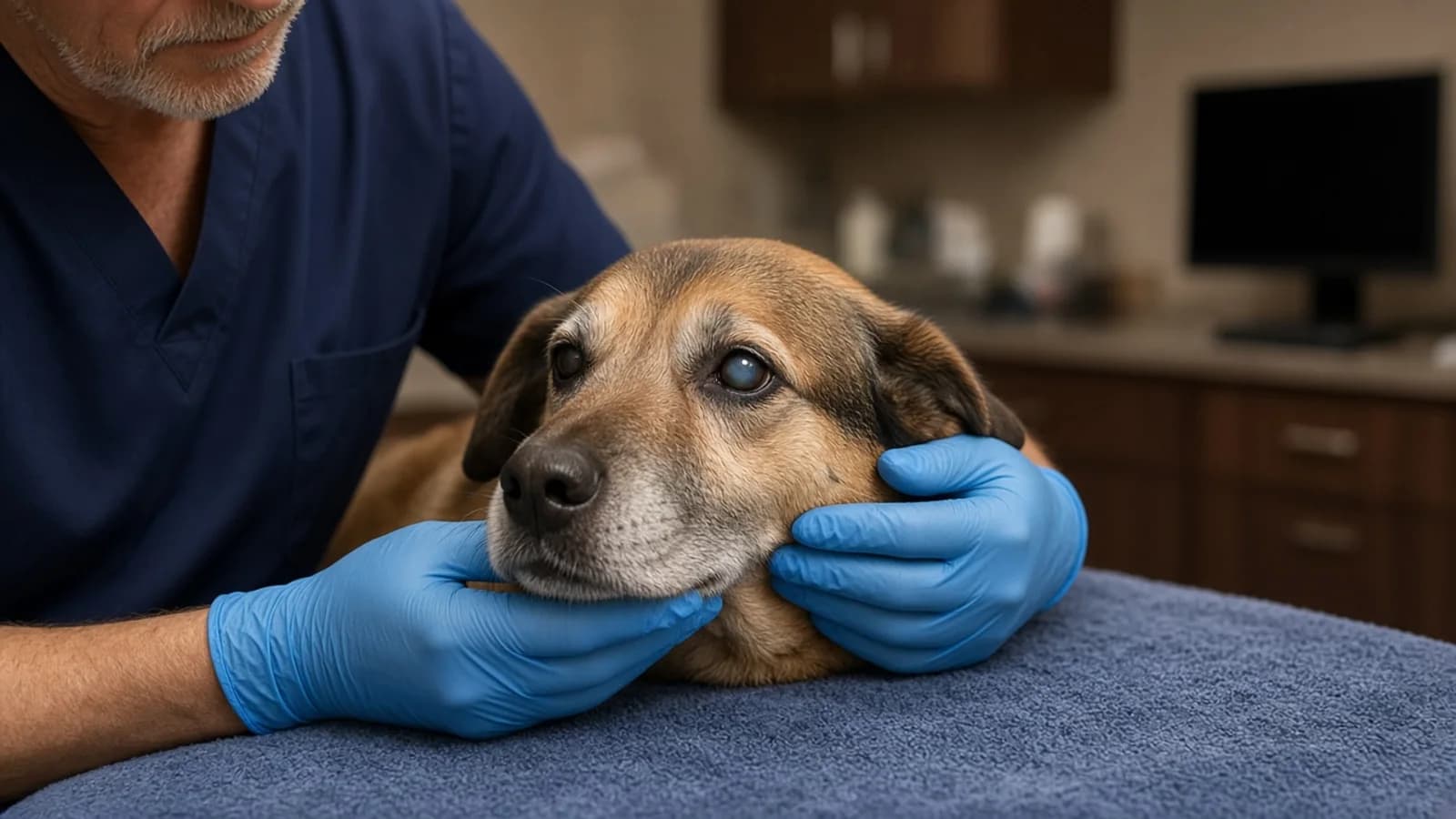
When a dog develops an advanced cataract, the lens turns cloudy, bluish-gray, or white. Scattered light destroys a focused image, so the dog's vision fails. The same opacity blocks the ophthalmologist's view: a slit lamp or an indirect ophthalmoscope cannot return a usable look at the retina, the retinal vessels, or the optic nerve. That is a failure of the examination, not a claim that no light at all reaches the retina. Enough unfocused light can still get through for an electrical test, which is why an ERG can be recorded after the view is gone.
That opacity creates the surgical problem. Modern canine cataract surgery, phacoemulsification, breaks up the cloudy lens with ultrasound and usually replaces it with an artificial intraocular lens. It is an elective procedure under general anesthesia, and the point of taking that risk is restoration of vision. Lens removal can restore vision only if the retina behind the cataract still responds. Cornell's Riney Canine Health Center states the boundary directly: if the retinas are not healthy, the dog will still be blind, or will become blind, even with surgery. In 2008 proceedings, Gwendolyn Lynch, DVM, DACVO, wrote that animals with a low-amplitude or flat ERG, or with a retinal detachment, usually are not candidates for cataract surgery. The electrical test is meant to find that out before anesthesia. It is not a promise about the result afterward.
Most veterinary ophthalmologists require a pre-operative electroretinogram, often with ocular ultrasound and sometimes gonioscopy, before cataract surgery. That is how Lynch summarized specialty practice in those 2008 proceedings. Cornell's ophthalmology service describes the same pairing inside a typical referral stay: an electroretinogram to demonstrate appropriate retinal function, and an ocular ultrasound to rule out retinal detachment. The American College of Veterinary Ophthalmologists recommends examination before a cataract is fully formed, because that is when the structures at the back of the eye, including the retina, can still be evaluated directly. Once the lens is opaque, the electrical test is how function is checked. A device maker, LKC Technologies, says an ERG must be performed on every cataract candidate. That sentence is a vendor recommendation, not a specialty-college rule. The reading of the tracing belongs to the ophthalmologist who examined the dog.
What an ERG Records, and Why It Works Through Dense Opacities
An electroretinogram is an electrical test of how the retina responds to light. VCA SouthPaws compares it with an electrocardiogram: an ECG records the heart's electrical wave, and an ERG records the wave produced by the retinal photoreceptors, the rods and cones. Drazek and colleagues, in a 2014 review in Veterinarni Medicina, state that full-field flash ERG is the most commonly used technique in veterinary ophthalmology for testing outer-retinal function, and that the ERG can detect hereditary retinal disorders before behavioral or ophthalmoscopic signs appear. VCA Advanced Veterinary Care Center adds a practical rule from its own service: on their setup, the larger the waveform, the better the retinal function.
Rods support vision in dim light. Cones support vision in daylight, including detail. VCA SouthPaws describes the ERG as the recorded response of those photoreceptors to light. The active sensor in the Lee 2009 montage sat on the cornea, with separate reference and ground sensors on the skin. The tracing is a sum of retinal electrical activity. It is not a picture of the fundus, and it is not a map of single cells.
Pet owners frequently ask how an electrical test can function through a cataract that has already blinded the pet. The answer lies in the fundamental physics of optics: the difference between image resolution and bulk photon illumination. To perceive a clear image—such as recognizing an owner's gesture or tracking a tennis ball—the cornea and crystalline lens must refract parallel light rays precisely onto corresponding focal points across the retinal surface. When cataractous lens fibers break down and precipitate proteins, light rays are scattered randomly in all directions, destroying image formation and rendering the dog functionally blind.
A focused image and a recordable retinal response are different physical events. Forming an image requires the lens to focus light. Triggering photoreceptors requires that enough light reach them. LKC Technologies states that even when a cataract or a corneal opacity affects vision, sufficient light still reaches a functional retina to generate an electrophysiologic response. That is the manufacturer's rationale for testing through a mature cataract. It is not a measurement of what percentage of light gets through, and it does not mean every opaque lens produces a normal-height tracing. The 2023 study below shows that some amplitudes fall as cataracts advance even when the authors' conclusion is that the retina may still be free of primary retinal disease.
Retinal Degeneration Can Hide Behind a Cataract
The requirement for pre-surgical electroretinography is rooted in real-world clinical data: in canine patients, neuroretinal disease frequently coexists with or directly precipitates cataract formation. Without an ERG, an ophthalmologist cannot differentiate a dog whose vision loss is caused solely by an isolated lens opacity from a dog whose vision loss is compounded by irreversible retinal decay.
How often retinal disease changes the surgery decision is documented in one hospital series, not as a national rate. Adkins and Hendrix (Journal of the American Animal Hospital Association, 2005) reviewed 244 dogs evaluated for cataracts at the University of Tennessee from January 2001 through December 2002. Fifty-four breeds were in that caseload. One hundred fifty-nine of the 244 dogs did not have cataract surgery. The most common reason was retinal degeneration, in 66 dogs. The abstract does not say how retinal degeneration was established in each dog, and a 2001–2002 referral caseload may not match current imaging or case mix. The count is still the clearest published reason this test exists: in that clinic, retinal degeneration was the leading recorded reason surgery did not proceed.
In the same series, cataract odds were significantly higher for six purebreds than for mixed-breed dogs. The abstract names them as the cocker spaniel, miniature schnauzer, toy poodle, Boston terrier, miniature poodle, and bichon frise. That is a finding about cataract odds in one university caseload. It is not a finding that every purebred has higher odds, and it is not, by itself, a list of breeds with progressive retinal atrophy.
Cataract odds and retinal-disease risk are not the same list. Eye Vet Clinic names cocker spaniels and miniature poodles among breeds affected by progressive retinal atrophy. LKC Technologies' clinical tables also list miniature schnauzer PRA, and prcd in toy and miniature poodles and in cocker spaniels, as inherited retinopathies in which the ERG can change before the ophthalmoscopic appearance does. The Royal Veterinary College describes PRA as progressive: signs noticed at night are likely to extend into daylight. Boston terriers and bichon frises are on the Adkins cataract-odds list. These sources do not put them on the PRA list.
Eye Vet Clinic's 2023 owner factsheet says most dogs with progressive retinal atrophy also develop cataracts, so the eyes look white over time, and that the cataract adds to the vision loss. Once a cataract has formed, the ophthalmologist can no longer examine the retina in detail, and an ERG can be used to look for PRA behind it. The Royal Veterinary College's fact file says cataracts may develop secondary to PRA and that a pet with PRA will most likely not be a candidate for cataract surgery. The factsheet also says a gene test on an oral swab is available for most breeds. A gene result shows hereditary status. It does not show whether the retina is producing a response on the day surgery is being considered. LKC's breed tables list prcd-PRA among the inherited retinopathies in which ERG changes can precede ophthalmoscopic and behavioral signs, including in cocker spaniels and in toy and miniature poodles. Those tables are a device maker's clinical summary, not a genetic-test menu.
Dogs with PRA have reduced or absent retinal responses on an ERG, the Eye Vet factsheet says, and surgical removal of the cataract does not stop vision loss when the retina is the cause. The same factsheet states a breed exception: in some breeds, especially cocker spaniels, cataracts can develop earlier than the retina fails, and surgery may prolong the time the dog remains sighted. All dogs with PRA are still expected to go blind. Whether a particular cocker spaniel has enough remaining retinal response for that conversation is the ophthalmologist's judgment after the ERG, not a rule an owner can apply from the breed name.
What Happens on the Day of the Test: Clinical Workflow
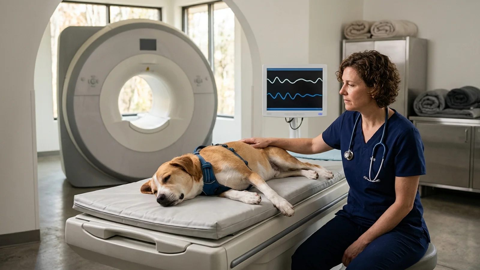
For pet owners, the word electroretinogram often raises questions about pain, restraint, and anesthesia. The Veterinary Eye Institute's client handout says the ERG and the ocular ultrasound are not painful and can be performed on most pets without sedation. The test records electrical responses from the eye. It is not a surgical incision. Whether a given dog is held awake, sedated, or, in a research recording, anesthetized depends on the hospital and is spelled out below from the pages that actually describe their own practice.
The diagram shows one common order of events. It is not a minute-by-minute protocol for every hospital.
graph TD
    A[Eye exam finds a cataract] --> B[Mydriatic drops, then dark adaptation]
    B --> C[Electrodes on the cornea and nearby skin]
    C --> D[Dark-adapted rod and mixed flashes]
    D --> E[Light-adapted cone and flicker flashes]
    E --> F[Compare the tracing with the ultrasound]
F --> G[Ophthalmologist discusses candidacy]The notes under the diagram are limited to what the cited hospital pages and papers actually describe.
Pharmacological Mydriasis (Pupil Dilation): Clinics dilate the pupil so the flash reaches a wider area of retina. There is no single drop strength to quote as the standard. In the Shih Tzu normal-values study by Lee and colleagues (Journal of Veterinary Science, 2009), the protocol used 0.5 percent tropicamide, applied twice and 15 minutes apart, followed by 20 minutes of dark adaptation. That concentration belongs to that study. Ask which drop and which waiting time your hospital uses.
Dark Adaptation (Scotopic Priming): Dark adaptation is the wait in the dark before the flashes, so rod photopigment can recover from room light. The Lee 2009 protocol used 20 minutes. VCA Advanced Veterinary Care Center says the test requires around 20 minutes of complete darkness. The Veterinary Eye Institute handout says the pet is placed in a dark exam room for 20 to 30 minutes. Those are three descriptions of a similar step, not one universal timer, and they are not something to reproduce at home.
Topical Corneal Anesthesia and Lubrication: The Lee 2009 recording was done under dim red light. The cornea was numbed with 0.5 percent proparacaine, and an artificial tear containing hydroxypropyl methylcellulose was applied before the contact-lens electrode. That is one published research montage. It does not mean every clinic uses those products, or that the eye is numb within a fixed number of seconds. The owner-facing claim that is actually printed is narrower: the Veterinary Eye Institute says the tests are not painful.
Three-Electrode Montage Placement: The team positions three lightweight sensors:
Active Electrode: In the Lee 2009 study the active sensor was an ERG-Jet contact-lens electrode resting on the cornea. Other hospitals use their own corneal electrodes, so the name on your report may differ.
Reference Electrode: In that same study the reference sensor was a needle electrode placed about midway between the lateral canthus, the outer corner of the eyelids, and the ear on the same side. It was not described as a fixed distance of a few millimeters from the eyelid. Some clinics use adhesive skin sensors instead. The report should name what was used.
Ground Electrode: In the Lee montage the ground needle was placed over the external occipital protuberance, on the midline at the back of the skull. Its role in that setup was to help the recorder handle electrical noise. Placement on another hospital's tracing may differ.
Scotopic Flash Recording: A fuller protocol then records dark-adapted responses: dimmer flashes aimed at rod function, then brighter flashes that mix rod and cone responses. Lee and colleagues recorded that kind of sequence on a portable mini-Ganzfeld unit. Wegg, Pollard, and Ofri (Veterinary Ophthalmology, 2023) used a full European College of Veterinary Ophthalmologists protocol with extensive rod and cone analysis in dogs presented for cataract surgery. A short screening flash, which some hospitals use only to ask whether the retina responds at all, is a different test from that full sequence. The report should say which one was done.
Light Adaptation and Photopic Recording: After the dark-adapted tracings, a fuller protocol records cone responses in the light, including single flashes and flicker. Wegg and colleagues reported cone-flicker amplitudes separately from single-flash cone responses, and they found cone-flicker amplitudes lower in mature and hypermature cataracts. Lee's handheld protocol also recorded a cone flicker response. Neither paper states a 10-minute light-adaptation rule or a 30-hertz flicker rate that every clinic must follow. Ask whether cone flicker was included.
Post-Test Inspection: The sensors come off when the recording is done. The owner pages cited here do not describe a universal rinse-and-stain step. If the cornea was numbed, ask the team whether they checked the corneal surface before discharge, and call them the same day if the eye is squinting or tearing.
Hospital Protocols on Restraint and Sedation: Hospitals do not share one restraint plan, and these pages should not be merged into a national rule. VCA Advanced Veterinary Care Center says the ERG can be performed on awake or sedated patients. The Veterinary Eye Institute handout says the tests are not painful and can be performed on most pets without sedation. Eye Vet Clinic's 2023 progressive-retinal-atrophy factsheet says sedation is often needed to carry out an ERG. The Lee 2009 normal-values study anesthetized healthy Shih Tzus with intravenous medetomidine and ketamine because it was a research recording, not because anesthesia is the pre-cataract standard. VCA SouthPaws explains why the test is ordered and does not say whether its patients are awake. Ask what this hospital does for a dog of this temperament.
ERG vs Ocular Ultrasound: Function vs Structure
When preparing for a cataract surgical evaluation, pet owners are frequently presented with a diagnostic plan that pairs an electroretinogram with an ocular ultrasound (B-scan ultrasonography). A sensible question often arises: 'If both tests examine the back of the eye, why do we need both? Doesn't the ERG tell us everything we need to know?'
The answer is that electroretinography and ocular ultrasonography evaluate completely distinct physiological domains. An ERG measures microscopic electrical function; an ocular ultrasound evaluates macroscopic physical structure. Relying on either modality alone creates an unacceptable diagnostic blind spot.
| Diagnostic Dimension | Electroretinography (ERG) | B-Scan Ocular Ultrasound |
|---|---|---|
| Primary Question Answered | Does the sensory retina possess viable electrical signaling function? | Is the retina physically attached and anatomically in place? |
| Diagnostic Modality | Electrophysiology (voltage potential over time) | Acoustic imaging (high-frequency sound wave reflection) |
| Primary Target Tissues | Rods and cones, recorded as the electrical waveform. VCA SouthPaws describes the ERG as a photoreceptor test. | Vitreous humor, retinal tissue sheet, choroid, retrobulbar orbit |
| Critical Pathology Detected | Reduced or absent responses when PRA is hidden by a cataract; a flat tracing is how VCA SouthPaws describes the SARDS diagnosis when the eye exam looks normal. | Retinal detachment and other posterior-segment structure. Cornell orders ultrasound to rule out retinal detachment before cataract surgery. |
| Key Diagnostic Blind Spot | Does not show whether the retina is physically detached, and does not test the optic nerve or the brain. | Cannot measure electrical life; an anatomically attached or severely degenerated retina can appear structurally normal while electrically dead |
| Patient Preparation & Time | Dark adaptation of about 20 minutes, or 20 to 30 minutes, depending on the hospital page, plus electrodes on and around the eye. Total appointment length is not one published number. | Images whether the retina is physically in place, including retinal detachment. It does not replace the electrical test and does not use the ERG's dark-adaptation step. |
| Surgical Candidacy Impact | A flat tracing usually means the dog is not a candidate for elective cataract surgery. A merely reduced amplitude is a specialist judgment, not an automatic stop. | A retinal detachment usually means the dog is not a candidate for routine cataract surgery. |
To appreciate why ophthalmologists require both tests, consider two contrasting clinical scenarios:
One pattern is advanced progressive retinal atrophy with the retina still lying in place. Ultrasound can look structurally unremarkable while the photoreceptors no longer respond. Eye Vet Clinic notes that dogs with PRA have reduced or absent ERG responses, and the Royal Veterinary College says a pet with PRA will most likely not be a candidate for cataract surgery. If only the ultrasound were used, that functional failure would be missed. This is an illustration of the gap between the two tests, not a case report.
The contrasting gap runs the other way. A retina can still produce some electrical signal and yet be physically detached, so an ERG alone would not show the detachment. Cornell orders ocular ultrasound to rule out retinal detachment before cataract surgery. Lynch's 2008 proceedings list retinal detachment, like a flat or low-amplitude ERG, as a usual reason not to proceed. Merck's retinal-detachment chapter, reviewed in 2024, calls detachment an important complication after lens surgery and lists B-scan ultrasonography among the tests used to find it. The two tests answer different questions, which is why they are booked together.
Together, the ERG and B-scan ultrasound provide an indispensable pairing: the ultrasound proves the movie screen is physically hanging on the wall, while the ERG proves the projector is plugged in and capable of displaying a picture.
Reading the Waveform: What Normal, Reduced, and Flat Mean
When the veterinary ophthalmologist reviews the ERG recording on the instrument monitor, they analyze a distinctive biphasic electrical tracing. Understanding what these peaks and troughs represent helps pet owners understand the clinical findings documented on the surgical evaluation sheet.
A standard full-field flash ERG waveform consists of two principal components:
The a-wave: The first downward deflection after the flash. Lee and colleagues measured a-wave implicit time from the stimulus to the trough. In their Shih Tzu series the mean was 15.12 milliseconds for the standard mixed rod-and-cone response, 13.42 milliseconds for the high-intensity mixed response, and 7.22 milliseconds for the cone response. A blanket '10 to 20 milliseconds' rule would misstate that cone result. Implicit time has to be read inside the protocol that produced it.
The b-wave: The large upward deflection that follows. In the Lee series, b-wave amplitude was measured from the a-wave trough to the b-wave peak. A taller wave is not automatically normal, and a shorter wave is not automatically retinal degeneration. Height is interpreted with cataract stage, age, and the device.
Ophthalmologists quantify this waveform using two primary measurements:
Amplitude (measured in microvolts, µV): The vertical voltage height of the wave. Lee and colleagues measured a-wave amplitude from the baseline to the trough, and b-wave amplitude from the a-wave trough to the b-wave peak. A taller wave is not a head count of living cells. Wegg and colleagues showed that some amplitudes also fall with advanced cataracts and with age.
Implicit Time (measured in milliseconds, ms): Implicit time is the delay from the flash to the peak of the wave. Wegg and colleagues found that rod implicit times did not change with advanced cataract stage even when some rod amplitudes did. Timing and height are separate measurements, and a longer implicit time is not, on that evidence, a shortcut diagnosis of early retinal disease.
Veterinary ophthalmologists group ERG results into three broad clinical categories:
Normal Waveforms: The tracing shows a- and b-waves under both dark-adapted and light-adapted conditions, judged against that device's own reference behavior rather than a universal cutoff. Lee and colleagues recorded a handheld mini-Ganzfeld ERG in 12 eyes of six healthy adult Shih Tzus under medetomidine-ketamine anesthesia. In that series, mean rod b-wave amplitudes ranged from 141.58 to 155.25 microvolts, the standard mixed rod-and-cone b-wave mean was 314.75 microvolts, and the high-intensity mixed b-wave mean was 329.42 microvolts. The cone b-wave mean was 37.75 microvolts, and the cone-flicker mean was 64.08 microvolts. Those figures are specific to that breed, device, and anesthetic protocol. They are not a pass mark for a cataract patient on another machine. A tracing the ophthalmologist calls adequate supports outer-retinal function. It does not, by itself, clear the dog for surgery.
Flat (Extinguished) Waveforms: The tracing is a flat line, with no clear a-wave or b-wave above the background noise. VCA SouthPaws describes the SARDS diagnosis as no photoreceptor response, a flat ERG, in a dog with sudden blindness and a normal-looking retinal exam. A flat tracing behind a cataract is the parallel finding for a retina that is not responding. Lynch's 2008 proceedings say animals with a flat or low-amplitude ERG usually are not candidates for cataract surgery, because removing the lens cannot restore vision if the retina is not responding. A low amplitude is a different sentence from a flat line. The next section is about why a reduced wave is not an automatic diagnosis.
Reduced (Attenuated) Waveforms: The waveform is present but displays diminished amplitudes or delayed implicit times. As explored below, an attenuated tracing does not automatically mean the dog has irreversible retinal disease; it represents an intermediate clinical picture that requires careful specialist interpretation.
What an ERG Cannot Tell You: Critical Limitations and Nuance
The electroretinogram is the test these specialty sources use to ask whether the outer retina responds before cataract surgery. It is not a complete prediction of vision. A reduced tracing and a normal tracing both still belong to the ophthalmologist who examined the dog.
Limitation 1: Reduced Amplitudes Do Not Automatically Mean Retinal Disease
A smaller wave is not, by itself, a diagnosis of retinal degeneration. The useful evidence is a retrospective pre-surgical series, not a slogan that every reduced tracing used to be called retinal disease.
Wegg, Pollard, and Ofri published that series in Veterinary Ophthalmology in 2023 (127 eyes of 67 dogs; the PubMed record is 35649104). Every dog had a full European College of Veterinary Ophthalmologists protocol, including extensive rod and cone analysis, before cataract surgery. The abstract, which is the part verified here because the full text is paywalled, supports the points below and does not support a single cutoff for 'too low to operate.'
Cataract Density Dampens Waveforms: Amplitudes, but not implicit times, of rod responses decreased with advanced cataracts. Single-flash rod amplitudes and rod-flicker amplitudes were significantly lower in eyes with mature cataracts, and single-flash rod amplitudes were also lower in hypermature cataracts. Cone-flicker amplitudes were significantly lower in mature and hypermature cataracts. Mixed single-flash rod-and-cone responses and cone responses were otherwise unaffected by cataract stage, except the mixed rod-and-cone a-wave amplitude in hypermature cataracts. The abstract does not say that every wave shrinks in proportion to how white the lens looks, and it does not prove the mechanism is light scatter. It says these particular amplitudes were lower.
Canine Aging Naturally Reduces Amplitudes: B-wave amplitude of the scotopic, mixed rod-and-cone, and photopic cone responses decreased with age by an average of 2.9, 7.5, and 1.5 microvolts per year, in that order (p < 0.01). The published abstract uses the word 'retrospectively' at that point; the three rates are listed in the same order as the three responses, and they should be read as those three age slopes, not as a second study design. They are average slopes in this population and protocol. They are not a calculator an owner can apply to one tracing.
The authors' conclusion is narrower than 'the cataract filtered the light, so the retina is healthy.' They wrote that lower ERG amplitudes in canine cataract patients may result from aging or from advanced cataracts and may not indicate retinal disease. An ophthalmologist who sees a reduced tracing in an older dog with a dense cataract has to judge which of those explanations fits. The paper does not authorize an automatic pass or an automatic stop.
Limitation 2: The ERG Evaluates the Outer Retina Only—Not the Optic Nerve or Brain
VCA SouthPaws describes the ERG as a test of the retinal photoreceptors, the rods and cones. It does not test whether the signal travels onward through the optic nerve to the brain. That is why a normal tracing cannot exclude central blindness.
VCA SouthPaws states the limit directly: a blind patient with a normal ERG and a normal eye exam most likely has central blindness, a problem in the brain or the optic nerve. LKC Technologies says ERG responses are extinguished in sudden acquired retinal degeneration syndrome and normal in optic neuritis. A normal tracing means the outer retina responded to the flash. It does not prove the signal reached the brain, and it does not prove the dog will see after cataract surgery.
Limitation 3: The Gap in Predictive Outcome Data
A Cornell research-award description says a complete cataract prevents visual evaluation of the retina, so the retina is routinely evaluated by ERG before surgery, and that there is a paucity of data on the correlation between the pre-operative ERG and post-operative visual function. That gap, the page says, prevents clear screening guidelines. The same page describes a planned study pairing pre-operative scotopic and photopic ERG with masked obstacle-course testing. Those results were not published on the page, so the award cannot be cited as proof that a given amplitude predicts how a dog will navigate after surgery. A normal ERG supports proceeding. It does not guarantee vision after surgery.
Limitation 4: A Normal Pre-Op ERG Does Not Prevent Post-Operative Complications
Passing a pre-operative ERG does not protect an eye from post-operative surgical hazards. Severe complications can arise after an otherwise flawless surgical procedure:
Post-Operative Retinal Detachment: Lynch's 2008 dvm360 proceedings describe post-operative retinal detachment as uncommon, at or under about 1 to 2 percent, and usually visually devastating. That percentage is hers, tied to a 2006 journal series she cites. It is not a rate printed in the Merck chapter. Merck's retinal-detachment chapter, reviewed in 2024, calls detachment an important complication after lens surgery and does not give this percentage. A normal pre-operative ERG does not prevent it. Cornell lists retinal detachment among post-operative complications alongside protracted inflammation, glaucoma, corneal ulcers, and infection, again without this percentage.
Post-Operative Glaucoma: Glaucoma is a separate risk from the ERG result. Lynch's 2008 proceedings put post-operative glaucoma risk under 10 percent for at least the first 3 years, higher in Boston terriers, cocker spaniels, and dogs with hypermature cataracts, and she uses gonioscopy alongside the ERG and ultrasound to assess the drainage angle. The ERG does not measure that angle or the eye pressure. Abnormal angle findings raise the glaucoma risk and, in her account, call for further client counseling rather than an automatic cancellation.
How the Results Shape the Surgical Decision
Once the electroretinogram and ocular ultrasound are completed, the veterinary ophthalmologist synthesizes the diagnostic findings alongside the patient's physical examination and systemic lab work to determine surgical candidacy.
Published Surgical Success Rates: Reported success is high only for selected eyes, and the two figures below are not the same statistic. The Merck Veterinary Manual says numerous studies have shown canine cataract surgery to be 90 to 95 percent successful within the first 1 to 2 years, with the best results before the cataract fully matures and before lens-induced uveitis is established. Cornell's Riney Canine Health Center reports success as high as 80 to 90 percent for ideal surgical candidates, and says that in general the sooner surgery is done, the better the outcome. Cornell also says the corneas must be healthy, and that if the retinas are not healthy the dog will still be blind, or will become blind, even with surgery. Neither range is a promise for an individual dog.
Why Early Evaluation Outperforms Waiting: Waiting for a cataract to 'ripen' is not what these sources recommend. In 2008 proceedings, Lynch called it a myth that cataracts need to mature before extraction and said earlier removal carries a better prognosis than chronic cataracts. Merck says results are best before the cataract fully matures and before lens-induced uveitis from leakage of lens material is established. Cornell ophthalmologist Kelly Knickelbein says the ideal time is before the cataract is mature, because mature and hypermature cataracts are more likely to cause lens-induced inflammation, lens instability, and loss of lens-capsule integrity. The American College of Veterinary Ophthalmologists recommends examination before the cataract is fully formed, so the retina can still be seen directly. Not every cataract progresses to blindness, which is another reason the exam comes before a surgery date.
The Diabetic Cataract Emergency: Diabetic dogs are a large share of cataract consultations, and two sources give figures that should not be averaged. Cornell's Riney Canine Health Center says about 75 to 80 percent of diabetic dogs develop cataracts within the first year of diagnosis, regardless of how well the diabetes is controlled. Those cataracts tend to form quickly and frequently cause severe lens-induced uveitis, which can result in glaucoma. The Merck Veterinary Manual's lens chapter reports diabetic cataracts in more than 85 percent of diabetic dogs regardless of diabetic regulation. It does not use Cornell's one-year window, and it says the lens can occasionally rupture and cause extensive uveitis. Both describe the same pathway: excess glucose in the lens is converted to sorbitol, which draws water in and swells the fibers. Lynch's 2008 proceedings add a timing caveat: diabetic cataracts can appear so fast that emergency surgery is considered, while elective surgery usually waits until diabetic control, such as a glucose curve or fructosamine, has been demonstrated. Prompt evaluation is the consistent advice. The diabetes workup itself is covered in our guide to Canine Diabetes Mellitus in Dogs.
Medical Management When Surgery Is Declined: A flat ERG, or retinal degeneration severe enough that the ophthalmologist declines surgery, ends the surgical plan. It does not end eye care. The Veterinary Eye Institute handout says an untreated cataract keeps leaking lens proteins, causing inflammation and predisposing the eye to lens luxation, retinal detachment, and glaucoma. Cornell's advice when surgery is not a fit is to discuss topical anti-inflammatory drops with the veterinarian and to have intraocular pressure checked about every four to six months. Merck says animals who do not have cataract surgery need chronic topical anti-inflammatory treatment and repeated pressure checks, because lens-induced uveitis can be followed by glaucoma. This guide does not name a drop or a dose. Cornell also notes that blind dogs generally adapt in a familiar home.
Questions to Ask Your Ophthalmologist and Records to Keep
Because canine ERG waveform amplitudes and implicit times depend directly on the instrument manufacturer, electrode type, and ambient room lighting, obtaining and preserving complete written documentation is essential for your pet's long-term ophthalmic history. If you ever seek a second opinion, relocate, or visit an emergency specialty clinic, having the original electrodiagnostic report prevents redundant testing.
Use this structured owner checklist during your cataract evaluation consultation:
Request the Full Graphical Waveform Report: Ask for a physical printout or digital PDF of the complete ERG summary. Ensure it includes the graphical waveform tracings showing the calibrated scotopic and photopic a- and b-waves, rather than just a brief text notation stating 'passed' or 'normal.'
Clarify the Specific Testing Protocol: Ask whether the recording was a short flash screen or a fuller rod, mixed rod-and-cone, and cone-flicker sequence. Wegg's group used a full European College of Veterinary Ophthalmologists protocol. Lee's group used the manufacturer protocol on a handheld mini-Ganzfeld. Knowing which family of test was done is what makes a later comparison possible. A note that only says 'passed' does not.
Verify Eye-Specific Lateralization (OD vs OS): Confirm that recordings are clearly separated for the right eye (OD - oculus dexter) and left eye (OS - oculus sinister). Cataract maturity, retinal degeneration, and electrical amplitudes frequently differ between the two eyes.
Review Concurrent Ultrasound and Gonioscopy Reports: Confirm whether a B-scan ocular ultrasound was performed to verify complete retinal attachment, and ask whether gonioscopy was performed to assess the iridocorneal drainage angles for glaucoma risk. Ensure both reports and sonogram printouts are included in your records.
Document Sedation or Anesthetic Agents: If sedation or anesthesia was used, ask for the drug names to be written in the discharge notes. The hospital pages cited here do not share one sedative, so the record should name whatever was actually given.
Maintain a Central Medical Record File: Keep your ophthalmic test reports filed alongside your dog's systemic blood panels, urinalyses, and cardiology screenings. For detailed guidance on organizing complex medical documentation across veterinary specialist visits, review our clinical framework on Veterinary Oncology First Appointment Records & Estimate Checklist.
Sources
1. American College of Veterinary Ophthalmologists (ACVO Public). Cataracts in Animals. Comprehensive owner guidance on cataract causes, surgery candidacy, and early ophthalmologist evaluation.
2. Cornell University College of Veterinary Medicine, Riney Canine Health Center. Canine Cataracts. Overview of canine cataracts, diabetes prevalence, pre-surgical ERG/ultrasound requirements, and surgical candidacy criteria.
3. Cornell University College of Veterinary Medicine. Evaluation of Electroretinography as a Predictor of Visual Function Following Cataract Surgery in Canine Patients. Research grant documentation addressing the scarcity of predictive outcome data linking pre-op ERG to post-op vision.
4. dvm360 / Gwendolyn Lynch, DVM, DACVO. Cataract Surgery in Veterinary Medicine Today (Proceedings 2008). Specialist continuing-education proceedings reviewing pre-surgical ERG/ultrasound/gonioscopy protocols, early surgery advantages, and retinal detachment risks.
5. Veterinary Ophthalmology / Wegg ML, Pollard D, Ofri R. Retrospective Evaluation of Pre-surgical Electroretinography Results in a Mixed-breed Canine Population Presented for Cataract Removal Surgery (2023). Peer-reviewed retrospective study demonstrating that rod amplitudes decline with cataract stage and b-wave amplitudes decline with age.
6. Journal of the American Animal Hospital Association / Adkins EA, Hendrix D. Outcomes of Dogs Presented for Cataract Evaluation: A Retrospective Study (2005). University teaching hospital study documenting that retinal degeneration was the primary reason for surgical rejection in 66 of 159 non-surgical dogs.
7. Journal of Veterinary Science / Lee JS, Kim KH, Jang HY, et al. The Normal Electroretinogram in Adult Healthy Shih Tzu Dogs Using the HMsERG (2009). Peer-reviewed clinical electrophysiology study detailing canine ERG electrode montages, ISCEV-derived protocols, and breed-specific reference amplitudes.
8. Veterinarni Medicina / Drazek M, Lew M, Lew S, Pomianowski A. Electroretinography in Dogs: A Review (2014). 2014 open-access review stating that full-field flash ERG is the most commonly used technique in veterinary ophthalmology for outer-retinal function, and that ERG can detect hereditary retinal disorders before behavioral and ophthalmoscopic signs.
9. Merck Veterinary Manual. The Lens in Animals. Professional reference on cataract staging, early surgical intervention, lens-induced uveitis, and 90–95% 1- to 2-year post-operative success rates.
10. Merck Veterinary Manual / Thomasy SM, DACVO. Retinal Detachment in Small Animals (Reviewed 2024). Clinical emergency reference on diagnostic testing for retinal detachment, B-scan ultrasonography, ERG indications, and post-operative complications.
11. VCA SouthPaws Veterinary Specialists & Emergency Center. Electroretinography. Hospital service documentation on the ECG analogy of ERG, pre-cataract testing, and differentiating retinal disease from central blindness.
12. VCA Advanced Veterinary Care Center. Electroretinogram (ERG) in Dogs & Cats. Clinical service guide on dark adaptation, electrode setup, awake versus sedated procedures, and waveform evaluation.
13. Veterinary Eye Institute. Cataracts and Cataract Surgery Client Guide (2023). Specialty practice client education flyer outlining pre-operative ERG and ultrasound pairing, dark adaptation, and long-term ocular risks.
14. Eye Vet Clinic. Progressive Retinal Atrophy (PRA) Factsheet (2023). Specialist clinic guidance on PRA behind cataracts, secondary cataract formation, breed variations, and DNA testing.
15. Royal Veterinary College. Progressive Retinal Atrophy (PRA) Fact File. Academic veterinary guide on PRA progression, secondary cataract development, and non-candidacy for cataract surgery.
16. LKC Technologies. Animal Clinical Applications of Electroretinography. Equipment manufacturer clinical overview explaining light penetration through mature cataracts and early electrophysiological detection of retinopathies.