Dog CT vs MRI: What Question Is the Specialist Trying to Answer?
Understand when dogs need a CT scan versus an MRI, how specialists choose by diagnostic question, anesthesia risks, what normal scans mean, and records to bring.

The Short Answer: Match the Machine to the Question
When a primary-care veterinarian or veterinary specialist advises that a dog needs advanced cross-sectional imaging, pet owners commonly ask which modality provides the superior view. The consensus among board-certified veterinary radiologists is unambiguous: neither CT nor MRI is universally superior. Instead, veterinary medicine selects the scanner that matches the specific tissue density, anatomical location, and clinical question under investigation.
Computed tomography (CT) is an X-ray-based modality that captures contiguous cross-sectional slices and three-dimensional volumetric reconstructions without the superimposition of overlying anatomy. The University of Tennessee teaching-hospital CT FAQ states that CT's sensitivity to subtle differences in x-ray attenuation is about ten times that of conventional radiography, which is why bone lysis and new bone can be seen earlier. The Merck Veterinary Manual adds that CT also has much better contrast resolution than standard radiographs and removes superimposition. That combination is why CT is the usual first study for dense bone, lung nodules, complex nasal and sinus anatomy, and vascular maps such as portosystemic shunts. 'Best' here means best for that question, not best in the abstract.
Magnetic resonance imaging (MRI) operates on different physical principles. Rather than using ionizing radiation, MRI places the patient in a powerful magnetic field—the Merck Veterinary Manual describes magnets up to 60,000 times as strong as the Earth's magnetic field—and uses radiofrequency pulses to perturb hydrogen protons in tissue. Because soft tissues differ in water content and in how those protons relax, MRI produces the soft-tissue contrast CT does not. Merck calls MRI the mainstay of small-animal neuroimaging for brain and spinal cord. It is the study used for intracranial disease, spinal-cord parenchyma, and disc material that is not mineralized. It is not a higher rank of the same test as CT.
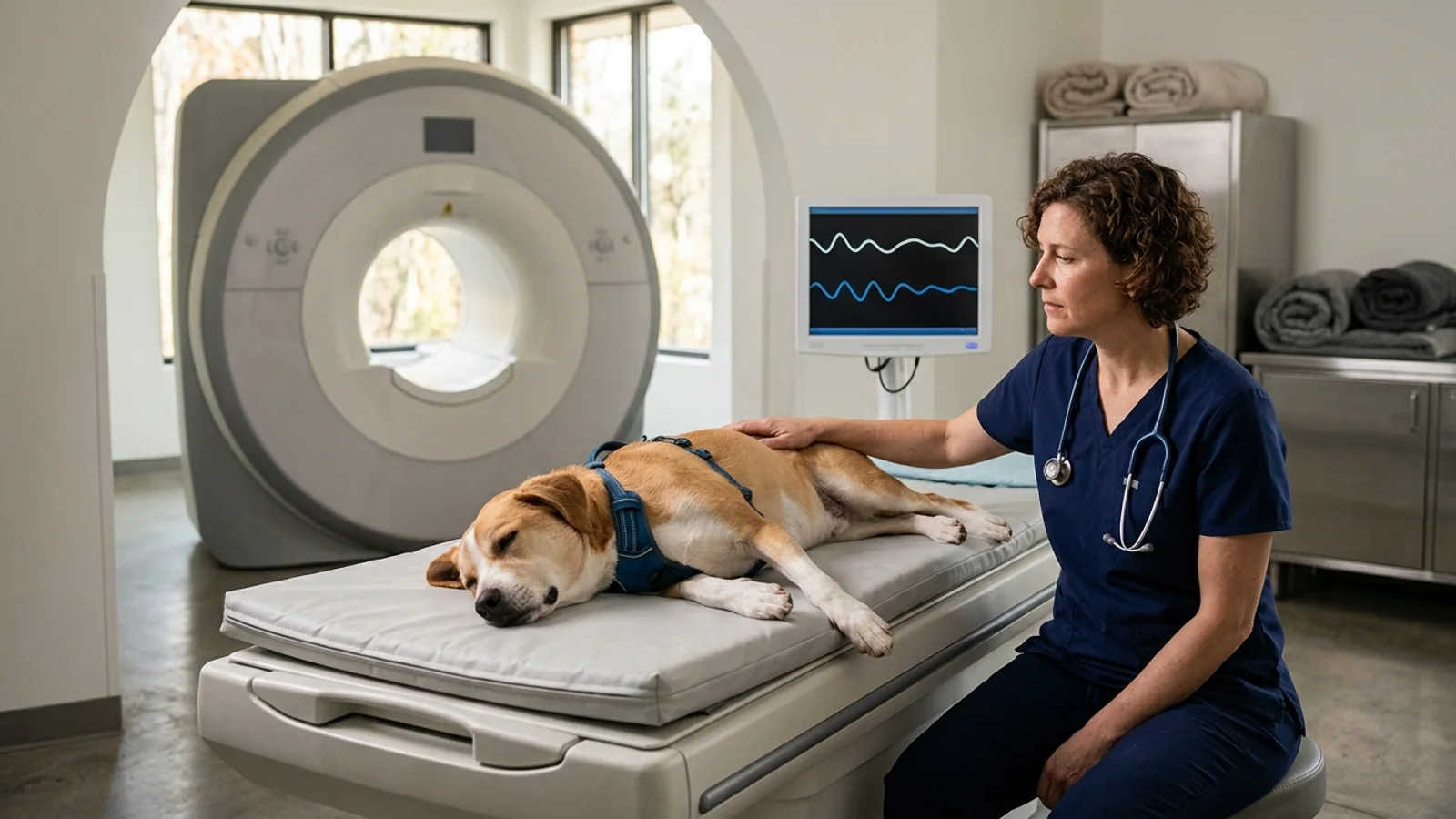
Veterinary patients cannot hold still for multi-minute sequences, so both studies require immobilization. Small-animal MRI is performed under general anesthesia because the sequences are long and motion-sensitive. CT is performed under general anesthesia or heavy sedation, depending on the region and the hospital's protocol. The controllable risk on the day is anesthetic fitness, not a ranking of the machines. The specialist chooses the scanner. The owner's job is the record packet, the bloodwork the hospital asked for, the fasting instructions that hospital gave, and a short list of questions.
What a CT Scan Answers Well in Dogs
Computed tomography excels whenever high spatial resolution, dense tissue differentiation, or rapid acquisition speed is essential. In a modern helical CT scanner, an X-ray tube rotates continuously around the patient while digital detectors capture projection data as the table moves smoothly through the gantry. The resulting volumetric datasets can be viewed in transverse, sagittal, and dorsal planes, or reconstructed into interactive 3D surface models.
In canine orthopedics and trauma, CT shows bone detail that overlapping radiographs hide. The University of Tennessee service calls CT the best osseous modality for complex fractures, for surgical planning of angular limb deformity, and for elbow dysplasia. It can show bone lysis and new bone earlier than radiographs. It does not publish a universal slice thickness, and it does not promise that every fragmented coronoid, ununited anconeal process, or osteochondrosis lesion will be visible before any radiographic change. For the limits of coronoid imaging, see elbow dysplasia.
For the lungs, the Merck Veterinary Manual states that metastatic lesions are far more evident at a smaller size on CT than on conventional radiographs, and that a CT done after nodules are already visible on radiographs will almost always find more nodules than the films show. Neither reference states a universal millimeter size at which a nodule becomes visible. That size-and-count advantage is why CT is used when the question is pulmonary metastasis. Separately, the Tennessee service calls CT angiography the gold standard for pulmonary thromboembolism, because it can show vascular filling defects that survey radiographs do not display. The protocol, including how many contrast phases are acquired, is the radiologist's choice.
Upper respiratory and cranial disease is another cornerstone of veterinary CT. When dogs develop chronic nasal discharge, persistent sneezing, facial asymmetry, or epistaxis, conventional skull radiography is a poor map. Merck states that skull CT is far more informative than radiographs because overlapping bone is removed. The Tennessee service adds that CT is extremely useful for nasal, orbital, and ear disease and can show very subtle bone lysis or proliferation. That map shows where to biopsy. It does not, by itself, separate fungal rhinitis from nasal neoplasia; tissue diagnosis still requires sampling and histopathology. If the question is whether disease has entered the brain, MRI remains the brain study. Middle- and inner-ear bone detail, including the tympanic bullae, is a CT strength for the same reason: the question is bone and air-filled spaces, not brain parenchyma.
In the abdomen, the Tennessee service calls CT the best modality for the morphology of vascular anomalies such as portosystemic shunts and for ectopic ureters, and a practical way to judge the origin and margins of abdominal masses. CT angiography is how those vessels are mapped for surgical planning. This page does not prescribe a dual-phase protocol or a particular occlusion device. The radiologist and surgeon choose both.
A practical advantage of CT is acquisition speed. Merck states that a modern scanner can image a small-animal abdomen or thorax in under 10 seconds, while positioning the patient often takes longer than the scan itself. Heavy sedation is an option the Tennessee service names alongside general anesthesia, when movement can be controlled that way. A thoracic study may still need a controlled breath-hold. That choice belongs to the anesthetist, not to a general rule about trauma patients.
Practice owners deciding whether to purchase a scanner, rather than which study one referred dog needs, are on a different question. The separate veterinary CT scanner buying guide covers equipment selection for a clinic. It does not choose CT versus MRI for one dog.
What CT cannot answer: Merck states that CT has essentially replaced myelography in small animals and that, for spinal-cord evaluation, CT remains relatively inferior to MRI. The Tennessee FAQ is more specific about discs: CT identifies mineralized disc material only. A non-mineralized extrusion can therefore look normal or nonspecific on CT, and the cord changes MRI is used to see are not a CT strength. That is a limit of the modality, not a list of every cord disease CT has been proven to miss.
What an MRI Answers Better
Magnetic resonance imaging is the mainstay of small-animal neuroimaging. It separates soft tissues by how hydrogen in water and lipid behaves in a magnetic field, which no X-ray method matches for brain and spinal cord. Different sequences emphasize different tissue properties. The radiologist chooses the sequence set for the question. A comparison of CT and MRI is not a protocol manual.
In veterinary neurology, MRI is the brain study. Washington State University's neurology service lists brain tumors, epilepsy, inflammatory brain disease, and stroke among the problems it evaluates with MRI and CT available. Merck's concrete comparison is narrower and more useful: a contrast-enhancing midbrain tumor would not be visible on routine skull radiographs and would likely be substantially underestimated on CT. Histologic names such as meningioma or glioma come from the pathologist or from the radiology report's differential, not from the choice of machine alone.
For spinal disorders, MRI provides direct, high-contrast visualization of the spinal cord parenchyma, epidural space, nerve roots, and intervertebral discs. In conditions such as IVDD in dogs, MRI can show non-mineralized disc material in the canal and the spinal cord itself. That is the gap CT leaves when the disc is not calcified. How bright the cord is on one sequence, and what that brightness means for recovery, is a reading of that dog's study. It is not a prognostic rule this comparison can state from the modality references. For cervical spondylomyelopathy (wobbler syndrome), MRI is the soft-tissue and cord study. The disease-specific page covers how disc-associated and bony forms differ. Bone-detail CT does not replace that cord assessment.
Outside the central nervous system, Merck notes that MRI is used for joints, muscles, cartilage, and ligaments, where tissue composition matters more than bone density. It does not establish that MRI can separate viable tumor from scar, necrosis, or edema in every mass. That distinction, when it can be made, is the radiologist's reading of a specific study.
MRI contrast agents are made for MRI and are different from the iodinated agents used for CT and radiography. Merck's example is a contrast-enhanced midbrain tumor that would not be visible on routine skull radiographs and would likely be substantially underestimated on CT. On safety, Merck says a link between these agents and chronic kidney disease has been described in some people and is not well documented in veterinary patients. The agents are still considered safe in animals, and they should be used with caution when kidney disease is already present. That is a caution, not a claim of minimal or zero kidney risk.
MRI has limits that matter when someone asks for it 'because it is more advanced.' Cortical bone contains little mobile hydrogen, and Merck states that MRI does not image cortical bone as well as CT, though it is useful for marrow and cartilage. Fine fracture margins and joint step-offs are CT questions. MRI is also extremely sensitive to ferromagnetic material. Merck notes that even the iron in an identity microchip can produce significant artifacts, that surgical stainless steel can still distort the image, and that a single steel pellet can make a study nondiagnostic. Radiographs of the area of interest are taken before the scan so the team can anticipate that artifact. Each sequence can take several minutes, and a brain or spine study uses several sequences, so magnet time is much longer than a CT acquisition. Total time under anesthesia also includes induction, positioning, and recovery. Neither reference converts that into one national appointment length.
Canine Advanced Imaging Decision Matrix: CT vs MRI by Diagnostic Question
The following comparison matrix illustrates how veterinary specialists match specific clinical questions to the appropriate imaging modality, highlighting the strengths and diagnostic boundaries of each scanner:
| Clinical Presentation / Anatomic Target | Primary Modality | Primary Diagnostic Question Answered | Limitations of the Alternative Modality |
|---|---|---|---|
| Complex Joint & Fracture Morphology (Pelvis, Elbow, Tarsus) | CT (Helical) | Fine cortical bone detail, articular incongruity, and the bone changes evaluated in elbow dysplasia and complex fractures. | MRI signal void in cortical bone makes fine osseous margin definition poor and prone to volume averaging. |
| Pulmonary Parenchymal Disease & Metastatic Staging | CT (Thoracic) | Finds metastatic lung nodules at a smaller size, and usually in greater number, than thoracic radiographs. A small-animal thorax can be acquired in under 10 seconds. | MRI acquisition is too slow; continuous respiratory and cardiac motion severely degrades lung parenchyma resolution. |
| Chronic Nasal Discharge, Sneezing & Sinus Asymmetry | CT (Skull) | Assesses fine nasal turbinate destruction, bone lysis, frontal sinus fluid, and cribriform plate integrity. | MRI poorly displays fine bone turbinates; however, MRI is added if tumor breaches the cribriform into the brain. |
| Intracranial Brain Disease (Seizures, Mentation Changes, Vestibular) | MRI (High-Field) | Exquisite soft-tissue detail of brain parenchyma, differentiating neoplasia, encephalitis (MUE), and infarction. | CT lacks soft-tissue contrast; misses non-calcified brain tumors, inflammatory encephalitis, and subtle edema. |
| Non-Mineralized Disc Extrusions & Spinal Cord Parenchyma | MRI (Spine) | Direct visualization of non-calcified disc herniation, intramedullary spinal cord edema, gliosis, and nerve roots. | CT detects only mineralized disc material; non-calcified disc extrusions in large-breed dogs remain invisible or inconclusive. |
| Mineralized Disc Extrusions in Chondrodystrophic Dogs | CT (Spine) | Rapid identification and surgical localization of dense, calcified disc material in the vertebral canal within minutes. | MRI provides equal or superior anatomical detail, but requires significantly longer anesthesia and higher procedural cost. |
| Vascular Anomalies (Portosystemic Shunts, Ectopic Ureters) | CT (Angiography) | Rapid contrast bolus tracking captures arterial and venous anatomy for surgical planning and shunt attenuation. | CT is the referenced best modality for shunt and ectopic-ureter morphology. MRI sequences take minutes, and motion limits many abdominal studies. |
Why One Symptom Can Point to Either Scanner
Dog owners are often perplexed when two dogs presenting with seemingly identical clinical signs—such as an inability to walk or sudden seizures—are referred for different imaging modalities. Understanding this scenario requires recognizing that veterinary specialists choose scanners based on the specific tissue pathology they suspect, not the superficial symptom.
The clearest example is acute hindlimb weakness. The Tennessee CT FAQ states that CT identifies only mineralized disc material, and that MRI remains the modality of choice in non-chondrodystrophic and large-breed dogs, which more often have discs that are not mineralized. A young dachshund or basset hound with acute spinal pain is the setting in which mineralized disc material is common, so CT can localize that mineral for surgery. The acquisition itself is short. Merck's figure for a small-animal thorax or abdomen is under 10 seconds of scanning, and positioning often takes longer than the scan. Neither reference states that the whole anesthetic lasts 15 minutes, or that decompressive surgery always follows in the same session. An older Labrador retriever or German shepherd with the same inability to walk is a different question: non-mineralized disc, a cord tumor, infection, or an infarct may all be in play, and none of those is reliably seen as mineral on CT. In that dog, spinal MRI is the study that shows the cord. Breed is a clue for the specialist, not a rule an owner uses to book a machine.
A similar decision tree governs the workup for canine seizures in dogs. When the question is seizures rather than a skull fracture, brain MRI is the study these references support. The Tennessee service says MRI remains the modality of choice for the brain, while CT remains useful when the question is bone, nasal passages, or the ears. Merck's contrast-MRI example is a midbrain tumor that routine skull radiographs would miss and that CT would likely substantially underestimate. Sequence-level seizure-focus measurements are read from that dog's images. They are not a result these references promise before the scan is read.
Acute vestibular signs (head tilt, circling, nystagmus) split the same way once a clinician has localized them. If that examination points to the ear, skull CT is the bone-and-bulla study: the Tennessee service calls CT extremely useful for ear disease and for subtle bone lysis or proliferation. If the examination points inside the skull, the question has moved to the brainstem or cerebellum, and MRI is the brain study. Those localizing signs are for the neurologist to assign. They are not a checklist an owner uses to book a scanner.
Anesthesia, Contrast, and Safety: Where the Real Risk Sits
When preparing a dog for an advanced scan, pet owners frequently worry about radiation exposure or scanner mechanics. However, in contemporary veterinary practice, the primary operational risk on the day of imaging is not the scanner itself, but the general anesthesia required to keep the patient motionless.
Motion ruins the images. Merck states that each MRI sequence can take several minutes and that small-animal MRI is performed under general anesthesia because of that motion sensitivity. The Tennessee service performs CT under general anesthesia or heavy sedation for the same reason. A thoracic CT may include a brief breath-hold so the lungs are not blurred; whether that requires general anesthesia is the anesthetist's decision for that dog, not a rule that every critical patient can be scanned sedated.
The numbers that exist are not imaging-specific. Shoop-Worrall and colleagues, writing in Veterinary Anaesthesia and Analgesia in 2022, studied 157,318 dogs with at least one general anesthetic or sedative event in UK primary-care practice. The events themselves were recorded from 2010 through 2013. They counted 159 deaths (0.10%) within 48 hours and 219 deaths (0.14%) within two weeks in which anesthesia or sedation could not reasonably be excluded as a contributor. Greater age, a poorer American Society of Anesthesiologists physical-status score, and more urgent procedures were associated with greater odds of death. In the same cohort, 8 of 89,852 dogs undergoing neuter surgery died in a way counted as anesthesia-related (0.009%). That gap is the point. The 0.10% figure is the all-procedure rate, about 1 in 1,000 dogs in that dataset, not the rate for a planned scan in a stable dog, and not a rate this paper measured for CT or MRI. A planned imaging visit is not the same population as an urgent procedure in a sick dog. The paper still does not report a CT or MRI death rate. Pre-anesthetic examination, the bloodwork the hospital requested, and monitored anesthesia are how the referral team manages the risk of whichever scan is chosen.
On radiation, the University of Tennessee radiology service says the dose to the patient is low, typically less than the dose for a person having a similar procedure, and that CT should be avoided in breeding animals during the first trimester of pregnancy. The service does not estimate cancer risk from one scan, and it does not tie any such risk to how long dogs live.
Iodinated contrast is routine for many soft-tissue CT studies. The Tennessee service says idiosyncratic reactions are extremely rare in animals. The complication it names as most common is contrast-induced kidney failure, which is still rare, is typically reversible with diuresis, and is more likely if the dog already has kidney disease or is dehydrated. Referral imaging requests commonly include a recent chemistry panel for that reason. A dog with known kidney disease should be discussed with the imaging service before contrast is planned. There is no single national rule, in these sources, that blood urea nitrogen and creatinine must be repeated inside a fixed 14-day or 30-day window. MRI contrast is a different drug. As Merck states, it is considered safe in animals and should be used with caution when kidney disease is already present.
The magnet hazard Merck describes is for ferromagnetic equipment brought into the room. A strong field can accelerate an object such as an oxygen cylinder to high speed, which is why those objects stay outside. Inside the patient, the manual's emphasis is image quality. A microchip can produce significant artifact. Surgical stainless steel has less ferromagnetic pull than iron, and it can still distort the picture. A steel pellet can ruin the study. Screening radiographs are how the team looks for that metal before the dog enters the room. A microchip is not a reason for an owner to cancel an MRI the specialist has recommended. These sources also do not call microchips or staples universally safe as projectiles. The hospital screens the patient and the room.
When the Scan Comes Back Normal
Few clinical experiences are more emotionally complex for a pet owner than investing significant emotional energy and financial resources into an advanced CT or MRI scan, only to have the specialist report: 'The scan is completely normal.' An owner's immediate reaction may be frustration, wondering if the procedure was a wasted effort. In clinical neurology and diagnostics, however, a normal scan is an exceptionally valuable, actionable finding.
The empirical check on 'the scan will find it' is a 2024 retrospective cohort from the University of Liverpool, published in Frontiers in Veterinary Science by Phillipps and Gonçalves. They included 412 dogs with two or more epileptic seizures and a normal inter-ictal neurological examination. Each had brain MRI on a 1 tesla or 1.5 tesla magnet (236 dogs at 1.5 T and 176 at 1 T). Sixteen dogs (3.9% as the abstract rounds 16/412, and 3.8% as the results section prints it) had a clinically significant structural cause, 13 of them suspected neoplasia. Seventy-six dogs (18.4% in the results; 18.5% in the abstract) had any MRI abnormality, and 60 of those 76 (78.9%) were judged incidental. The incidental findings listed most often were Chiari-like malformation (30), otitis (20, including 15 middle-ear and 5 external-ear cases), and syringohydromyelia (13). Ventricular dilation was recorded in 6. The authors did not treat those findings as the cause of the seizures.
Furthermore, the Liverpool study established that the probability of identifying a structural brain lesion is intimately dependent on the dog's age at seizure onset:
First seizure before 6 months of age: 0% structural lesions (0 of 25 dogs).
First seizure from 6 months until the first birthday: 0% structural lesions (0 of 41 dogs).
First seizure from 1 year through 6 years: 1.6% structural lesions (4 of 256 dogs).
First seizure after 6 years through 8 years: 5.9% structural lesions (3 of 51 dogs).
First seizure after 8 years of age: 23.1% structural lesions (9 of 39 dogs).
Those percentages do not cancel the International Veterinary Epilepsy Task Force list, and they also do not prove the list should be applied to every dog. De Risio and colleagues, in BMC Veterinary Research (2015), recommend brain MRI with a veterinary epilepsy protocol, plus routine cerebrospinal fluid analysis, after reactive seizures have been excluded, when any of the following is present: onset before 6 months or after 6 years of age; inter-ictal neurological abnormalities consistent with an intracranial localization; status epilepticus or cluster seizures; or a previous presumptive diagnosis of idiopathic epilepsy that is drug-resistant to one antiepileptic drug titrated to the highest tolerable dose. The Liverpool dogs all had a normal inter-ictal examination, which already removes one of those triggers. Inside that narrower group, structural lesions were uncommon, including zero of 25 dogs with onset before 6 months, and they rose to 9 of 39 (23.1%) only when the first seizure was after 8 years of age. The authors' conclusion is to decide case by case. It is not 'skip the MRI the neurologist recommended,' and it is not 'a normal scan proves the diagnosis.'
Idiopathic epilepsy is a diagnosis of exclusion. The IVETF text says the diagnosis rests on onset age, a normal inter-ictal examination, and exclusion of metabolic, toxic, and structural brain disease. The same paper splits confidence into tiers. Tier I uses two or more unprovoked seizures at least 24 hours apart, onset between 6 months and 6 years, a normal examination, and an unremarkable minimum blood and urine database. MRI is not required for that tier. Tier II adds a normal epilepsy-protocol brain MRI and routine CSF analysis. A normal MRI therefore supports a higher-confidence exclusion of a structural cause. It does not by itself prove idiopathic epilepsy, and the consensus does not say a clinician cannot counsel an owner or adjust medication unless MRI has been performed. The Liverpool study also did not sample CSF in every dog (274 of 412 had CSF collected) and did not perform EEG, so a normal MRI in that paper was not a complete workup.
The same honesty applies outside the brain. A normal spinal MRI makes a large compressive disc extrusion, an obvious cord mass, or a destructive vertebral infection less likely, and it often moves the plan away from immediate decompression. It does not definitively exclude every cause of back pain. Some disc material is still hard to see, some infections and tumors need CSF, culture, or biopsy, and some pain is in muscle or joints outside the field. What a normal study ruled out is a sentence in that dog's report, not a universal exclusion list.
The Owner's Checklist: Records, Bloodwork, and Videos Before the Scan
While the veterinary radiology team manages scanner physics, anesthesia protocols, and image post-processing, the dog's owner controls the preparation that ensures a smooth, safe, and diagnostic procedure. Specialty hospitals operate most efficiently when referral records and clinical documentation are complete prior to arrival.
Follow this structured preparation checklist before your dog's imaging appointment:
Primary Veterinary Referral and Imaging Request Form: Specialty practices and academic teaching hospitals require a formal referral from your primary-care veterinarian. The referring doctor completes an imaging request form specifying the anatomical region of interest, the clinical history, and the requested study (CT versus MRI). Confirm with your primary clinic that this paperwork has been submitted.
Complete Medical Records and Prior Imaging Data: Gulf Coast Veterinary Specialists describes the usual packet as recent exam notes, laboratory results, imaging reports, and a summary of treatments already tried, sent by the primary veterinarian. Ask what that hospital wants on file before arrival. These sources do not set a 24-hour or 48-hour deadline. If survey radiographs or dog ultrasound examinations were performed, have the digital DICOM files transferred directly so the radiologist can compare baseline anatomical structures.
Recent Pre-Anesthetic Bloodwork: Scissortail Veterinary Specialists asks referring veterinarians for recent blood work, a complete blood count and a chemistry panel, with the imaging request. How recent that panel must be is a hospital rule, and it changes if the dog is acutely ill or already has kidney disease. These sources do not authorize a homemade 14-to-30-day window.
Detailed Medication and Supplement Inventory: Bring a list of prescription medicines, preventives, pain medicines, and supplements, or photographs of the labels, including the dose and how often each is given. Do not change the morning doses on your own. The clinical team will say what to give and what to hold.
Smartphone Video Documentation of Intermittent Episodes: If your dog is being evaluated for seizures, episodic stumbling, paroxysmal dyskinesias, lameness, or collapse, capture high-definition video of the episodes. Dogs rarely demonstrate transient neurological or orthopedic signs during a brief consultation, and video recordings provide the specialist with invaluable diagnostic clues for neuro-localization.
Adherence to Facility-Specific Fasting Instructions: Fasting instructions are given when the appointment is scheduled. Anesthesia blunts the reflexes that protect the airway, so a full stomach raises the chance of aspiration. Follow that hospital's written instructions. Do not substitute 'nothing after midnight,' an 8-to-12-hour fast, or a personal decision to pull the water bowl. Puppies and diabetic dogs often have different instructions, and only the clinic scheduling the anesthetic should set them.
Closed-Loop Radiologist Reporting: Following scan completion, the cross-sectional image series is reviewed by a board-certified veterinary radiologist (Diplomate of the American or European College of Veterinary Radiology). The radiologist generates a comprehensive formal report that is sent directly to your primary veterinarian and attending specialist, who will interpret the findings and coordinate subsequent medical or surgical care.
Questions to Ask Before You Schedule — and What a Referral Really Means
Receiving a recommendation for a veterinary specialist consultation and advanced imaging can be an intimidating milestone for any pet owner. However, a referral does not automatically indicate a grave or untreatable diagnosis. As specialty clinicians emphasize, a referral simply reflects modern veterinary medicine matching a complex diagnostic question to specialized technology, dedicated anesthesia personnel, and board-certified expertise.
Before scheduling your dog's scan, bring these targeted questions to your consultation to ensure complete clarity regarding procedural goals, safety safeguards, and cost expectations:
What specific clinical or surgical question will this scan answer that previous physical exams, blood tests, or survey radiographs could not resolve?
Why is this specific modality (CT versus MRI) preferred for my dog's symptoms, and could the alternative modality provide equivalent or superior information?
If the scan reveals the suspected surgical or medical condition, what are our immediate treatment options, and what is the expected long-term prognosis?
What diagnostic steps or medical management pathways will we follow if the scan returns entirely normal?
What individualized anesthesia and monitoring protocols are planned for my dog, taking into account their age, breed, and baseline bloodwork?
Does the written estimate cover the pre-anesthetic exam, anesthesia medications, intravenous contrast agents, recovery monitoring, and the radiologist interpretation fee?
When will our primary-care veterinarian receive the official radiologist report, and who will oversee ongoing medical management?
By focusing on the diagnostic question behind the scan, understanding the respective strengths of CT and MRI, and preparing thorough medical records, pet owners and veterinary teams can navigate advanced imaging with clarity, confidence, and compassion.
Sources
The clinical recommendations, diagnostic criteria, and epidemiological statistics cited in this guide are derived from the following peer-reviewed veterinary literature and authoritative professional references:
Merck Veterinary Manual. Computed Tomography in Animals. Professional reference on veterinary CT: cross-sectional slices without superimposition, better contrast resolution than radiographs, metastatic lung nodules seen smaller and usually more numerous than on radiographs, and a full small-animal thorax or abdomen acquired in under 10 seconds. Reviewed August 2024, updated April 2025.
Merck Veterinary Manual. Magnetic Resonance Imaging in Animals. Professional reference on veterinary MRI: no ionizing radiation, mainstay of small-animal brain and spinal-cord imaging, cortical bone seen less well than on CT, contrast agents different from CT contrast, and metal artifact including microchips. Reviewed August 2024, updated April 2025.
University of Tennessee College of Veterinary Medicine, Veterinary Medical Center Radiology. CT FAQ. One teaching hospital's CT FAQ: about tenfold sensitivity to subtle attenuation differences versus radiographs, CT for bone, lung, nasal and ear disease, shunt morphology, and mineralized disc material only, plus radiation and iodinated-contrast cautions. It describes that hospital's practice, not a national standard.
De Risio L, Bhatti S, Muñana K, et al. International Veterinary Epilepsy Task Force consensus proposal: diagnostic approach to epilepsy in dogs. BMC Veterinary Research 2015;11:148. Consensus guidelines defining age parameters, clinical indicators, and standardized protocols for brain MRI and CSF analysis in canine epilepsy workups.
Phillipps SA, Gonçalves R. High-field MRI findings in epileptic dogs with a normal inter-ictal neurological examination. Frontiers in Veterinary Science 2024. Retrospective cohort study of 412 dogs analyzing structural lesion yield, age-stratified prevalence, and incidental findings on high-field brain MRI.
Shoop-Worrall SJW, O'Neill DG, Viscasillas J, Brodbelt DC. Mortality related to general anaesthesia and sedation in dogs under UK primary veterinary care. Veterinary Anaesthesia and Analgesia 2022;49(5):433-442. Large-scale epidemiological investigation of 157,318 dogs establishing population-level 48-hour and 2-week anesthesia mortality rates and risk factors.
Washington State University Veterinary Teaching Hospital. Neurology Service. Academic clinical service overview detailing veterinary referral requirements, advanced multi-modality CT and MRI imaging, and specialist diagnostic workflows.
Scissortail Veterinary Specialists. Imaging Referrals. Workflow documentation outlining veterinary referral protocols, pre-anesthetic screening laboratory requirements, patient fasting instructions, and board-certified radiologist reporting.
Gulf Coast Veterinary Specialists. Your First Visit to a Veterinary Specialist: What to Bring + How Referrals Work. Client guidance detailing the veterinary referral process, medical records preparation, symptom timelines, and episode video documentation.