Equine PSSM1 Testing: A Negative Result Does Not Exclude Muscle Disease
Understand what equine PSSM1 and muscle-panel results can establish, why P2–P4 markers lack diagnostic validation, and when vets consider CK testing or biopsy.

The short answer: a panel covers specified variants
A valid negative genetic test for the established GYS1 variant excludes type 1 polysaccharide storage myopathy (PSSM1). It does not exclude PSSM2, myofibrillar myopathy (MFM), recurrent exertional rhabdomyolysis, or other causes of muscle pain and atrophy. If GYS1 is only one component of a larger panel, each additional result needs its own interpretation. The number of tests bundled together does not establish how completely the panel covers a horse's clinical problem. PSSM1 research overview; Equine muscle-disease categories.
A positive GYS1 result establishes the PSSM1 genotype and is clinically useful. It still does not prove that every episode of stiffness, poor performance, or muscle loss is caused by that variant. A veterinarian must connect the finding to the horse's history and examination. This distinction preserves the value of validated genetic testing while avoiding a diagnosis that overlooks a second problem.
Commercial P2, P3, and P4 markers offered for PSSM2 or MFM need a different interpretation: the studies discussed below did not validate them as diagnostic markers in the populations examined. A positive or negative result for those markers cannot replace a clinical workup. Warmblood and Arabian validation study; Quarter Horse validation study.
What a PSSM1 result can and cannot tell you
PSSM1 is associated with a dominantly inherited variant in GYS1, the gene encoding glycogen synthase 1. The University of Minnesota links this variant to abnormal sugar accumulation in skeletal muscle. Genetic testing can use blood or hair-root DNA, according to the Merck reference. This is a defined gene–disease relationship, rather than a general search for anything that might cause tying-up. University of Minnesota PSSM1 research; PSSM1 diagnosis in the clinical reference.
Dominant inheritance means a horse with one copy can be affected; it does not mean every horse with that genotype has the same signs or severity. Utrecht's scientific summary describes differences in expression between horses with one and two copies and notes the influence of management. Its broad statement about the usefulness of PSSM1 testing should not be converted into a laboratory-specific percentage for analytical specificity. Utrecht scientific summary, 2019.
For a report review, separate three questions: Was the established GYS1 variant detected? Does that establish the PSSM1 genotype? Does PSSM1 explain the presenting episode? The first two concern the genetic finding; the third requires clinical interpretation. An accurate report can answer the genetic question without resolving every cause of the horse's signs.
Minnesota also reports that a second variant in RYR1 can worsen PSSM signs in some individuals. That is a reason to interpret relevant findings together, not to assume that every GYS1-positive horse has an additional variant or a severe outcome. Genetic modifier described by Minnesota.
Why a negative panel does not clear every muscle disease
The Merck overview groups equine muscle disorders into exertional, nonexertional, and other presentations. Its differential includes nutritional, toxic, inflammatory, traumatic, and inherited causes. PSSM1 is one part of that range. A DNA assay directed at one inherited variant cannot answer whether a horse has acquired muscle injury or a different disorder. Differential diagnoses of equine myopathies.
PSSM2 and MFM also illustrate why a normal muscle-enzyme result is not universal clearance: Merck lists exertional myopathies that can occur with normal CK activity. A negative DNA result and a normal CK result answer different questions. Neither should be stretched into a statement that the horse has no muscle disease.
The practical question after a negative result is therefore whether the remaining clinical findings have been explained. Repeating a broader-looking panel is useful only if an additional, validated target addresses an unresolved question. More reported variants do not automatically provide more relevant evidence. The veterinarian may instead need to reassess the history, examination, laboratory findings, or the possibility of a problem outside muscle.
PSSM2 is a biopsy-based classification, not one DNA diagnosis
PSSM2 describes horses with abnormal muscle polysaccharide findings that lack the PSSM1 GYS1 variant. Minnesota's older research summary reports that about 25% of its described Quarter Horse PSSM cases were not due to GYS1, and that the variant had not been found in at least 18 breeds previously reported to have PSSM. These are observations about described cases and breeds, not a prevalence estimate for all horses or a reason to skip testing in an individual. University of Minnesota PSSM2 research summary.
The term can encompass more than one underlying disorder. It should not be treated as the name of a single established mutation. Failure to validate proposed markers also does not establish that every condition grouped under PSSM2 lacks a genetic contribution. Those are separate scientific questions.
What the P2–P4 studies actually found
The Quarter Horse study included 229 healthy controls and 163 horses with biopsy-defined PSSM2, totaling 392 Quarter Horses and related stock breeds. One or more P variants occurred in 57% of controls and 61% of PSSM2 cases. The authors found no significant association between the tested markers and PSSM2. They estimated that using those markers diagnostically would misclassify 57% of healthy controls and miss about 40% of biopsy-confirmed cases. The study appeared online in 2022 and in a 2023 journal issue. Quarter Horse P2–P4 study.
The separate Warmblood and Arabian investigation compared 54 Warmblood controls with 68 PSSM2/MFM cases and 30 Arabian controls with 30 cases. It likewise found no significant association for the commercial markers studied. The authors did not support their use for myopathy diagnosis, prepurchase examinations, or selection decisions. The paper appeared online in 2020, with a 2021 journal issue. Warmblood and Arabian P2–P4 study.
These findings concern P2, P3, and P4, including the P3 variants specified in the papers, in those populations. They are not a measured failure rate for every breed, every marker, or a future test. Nor does finding a variant in healthy horses, by itself, prove that the variant has no biological effects. The diagnostic problem is the lack of demonstrated correspondence with the disease being claimed.
A laboratory's ability to detect a DNA sequence accurately is different from evidence that the sequence predicts a clinical disorder. That distinction explains how a report can correctly identify a P variant yet fail to establish PSSM2. It also explains why a negative report for those markers cannot exclude biopsy-defined disease.
Variant coverage versus the clinical question
Use this comparison to frame a discussion with the veterinarian and laboratory. It is a synthesis of the cited evidence, not a treatment algorithm or a list of tests every horse needs. Cornell lists genetic testing for selected equine myopathies; the validation papers explain why the proposed PSSM2 markers need separate scrutiny. Cornell Equine Myopathy Plan; P2–P4 validation in Warmbloods and Arabians.
| Clinical question | Test or target | What a positive result supports | Limit of a negative result | Question for the veterinarian |
|---|---|---|---|---|
| Does this horse have the PSSM1 genotype? | Established GYS1 variant | The genetic basis for PSSM1; current signs still need interpretation. | With a valid sample and assay, excludes PSSM1, not other myopathies. | Does PSSM1 account for the current findings? |
| Do proposed markers establish PSSM2 or MFM? | Commercial P2, P3, P4 markers studied in the cited papers | Presence of the assayed markers; not a validated PSSM2/MFM diagnosis in the studied populations. | Does not exclude PSSM2 or MFM. | What clinical and biopsy evidence supports the diagnosis? |
| Could MYH1-associated disease explain muscle atrophy? | MYH1 E321G for MYHM | A genotype associated with MYHM, with variable clinical expression. | Absence of this variant does not exclude other causes of atrophy or muscle injury. | Are the signs compatible with MYHM or another cause? |
| Is the established MH variant present? | RYR1 variant for malignant hyperthermia | Inherited susceptibility relevant to the clinical and anesthesia history. | Addresses the assayed variant, not every cause of hyperthermia or rhabdomyolysis. | What does this finding mean for this horse's care? |
| Is HYPP relevant to the presenting problem? | A specific HYPP genetic test | The reported HYPP genotype, interpreted for that condition. | Does not exclude other muscle disorders or causes of weakness. | Does this test address the actual clinical presentation? |
| Is a GBED result answering the right question? | A specific GBED genetic test | The reported genotype for a condition Cornell identifies in foals. | Does not clear an adult horse of causes of tying-up. | Is the purpose a foal-disease question or a separate genetic assessment? |
The negative-result column is deliberately narrow. A report should identify what was actually assayed before anyone describes a condition as excluded. If the laboratory renames a panel or changes its contents, compare the variant lists rather than assuming that two panels with similar names have identical coverage.
When CK, nutritional tests, or muscle biopsy add evidence
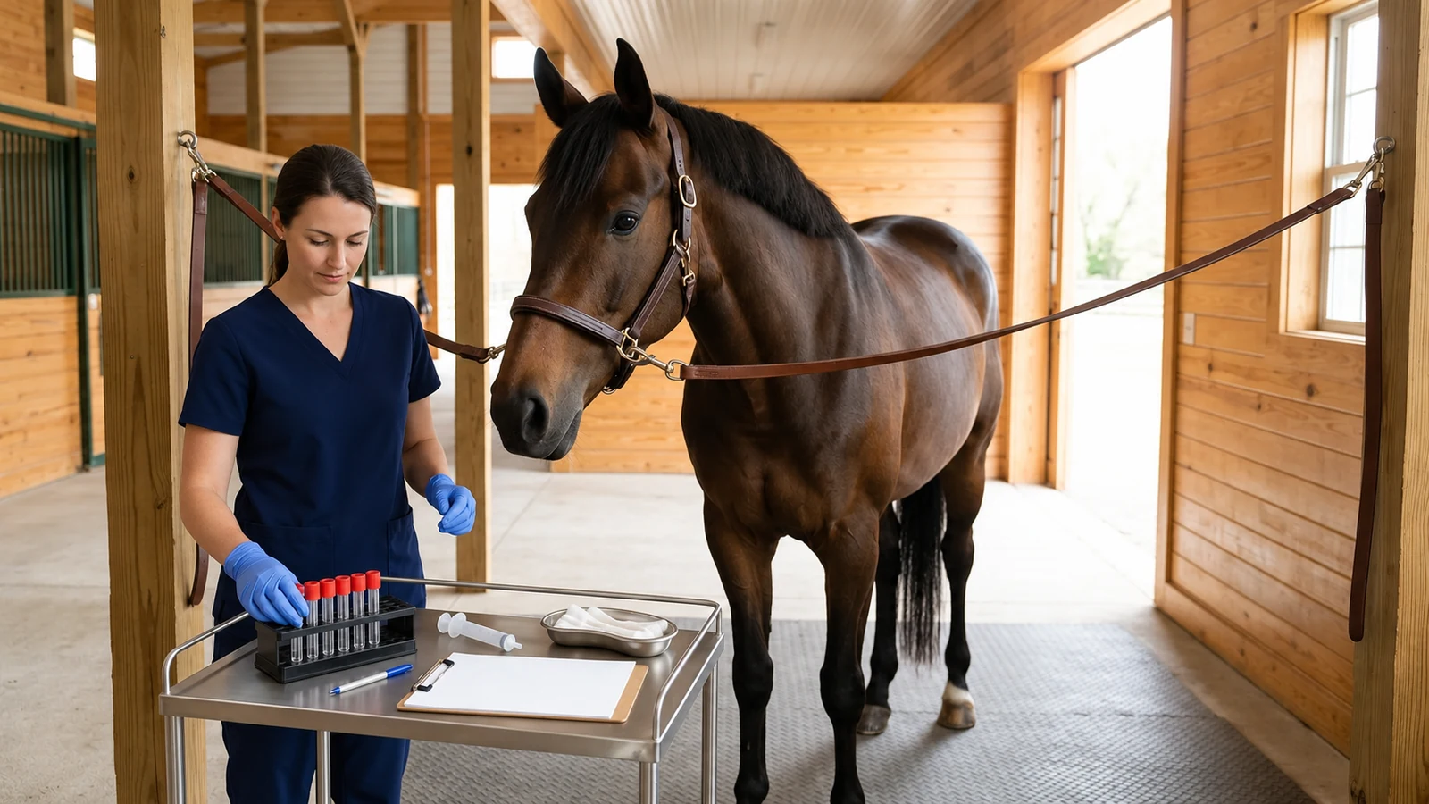
Cornell's Equine Myopathy Plan includes CK testing associated with exercise, whole-blood selenium, serum vitamin E, and referral guidance for muscle biopsy. Its notes emphasize that biopsy recommendations vary with the suspected disorder. This is a laboratory plan for clinician-directed evaluation, not a universal sequence that starts only after every DNA test is negative. Cornell laboratory plan and notes.
The useful distinction is between inherited sequence information, evidence about current muscle injury, nutritional findings, and tissue findings. These can complement one another. A veterinarian chooses which question matters at a particular point; a positive genetic result does not automatically make all other assessment unnecessary. Conversely, an unexplained clinical problem does not establish a genetic cause simply because the first laboratory results are unremarkable.
For suspected PSSM2, Merck describes diagnosis using abnormal muscle glycogen storage together with a negative GYS1 result. Biopsy can also help distinguish other myopathies. Its value depends on the clinical question and the receiving laboratory's requirements. It should not be presented as an infallible final answer for every horse. Clinical interpretation of muscle biopsy.
Utrecht notes that a young, untrained horse can have a normal biopsy before abnormal storage becomes visible. The Quarter Horse paper also identifies delayed shipping of fresh muscle as a potential source of false-negative PSSM2 findings. Neither observation supports a universal age cutoff or a fixed shipping-time threshold. Specimen planning and interpretation belong with the veterinarian and diagnostic laboratory. Biopsy limitations in the Utrecht summary; Sample-delay limitation in the Quarter Horse study.
Merck specifically advises against using muscle biopsy for prepurchase examinations or breeding decisions. A procedure useful in a symptomatic horse is not automatically an appropriate screening test for a different decision. Ask what the result could change before pursuing it.
MYH1 and the other panel entries answer separate questions
MYH1 E321G is associated with myosin heavy chain myopathy (MYHM), including immune-mediated myositis and nonexertional rhabdomyolysis in Quarter Horse-related breeds. It is a different target from GYS1. A negative PSSM1 result says nothing about whether MYH1 was tested; check the actual report. MYH1 clinical-expression study.
In the 2022 retrospective owner-questionnaire study, muscle atrophy was reported in 17 of 100 heterozygous horses (17%) and 8 of 10 homozygous horses (80%). It also occurred in 29 of 275 horses without the variant (11%). The small homozygous group, selected respondents, and owner-reported histories limit how those percentages apply to an individual horse. They are study observations, not a personal forecast or population-wide lifetime risk.
The study illustrates both incomplete expression of a variant and the existence of muscle atrophy in horses without it. A laboratory finding can inform the veterinarian's suspicion while leaving other explanations open. For HYPP, GBED, and MH, the same discipline applies: read each entry as a condition-specific result. Being printed beside PSSM1 does not make another test a substitute for PSSM1 testing, or make the panel a complete explanation of weakness.
A five-step review of the evidence behind a panel result
The following is a report-review framework derived from the validation studies. It helps keep the test's claim aligned with its evidence; it does not determine an individual horse's diagnosis. Diagnostic-validation methods and conclusions.
Identify the exact target. Record the gene, variant, reported genotype, and panel contents. A condition name or a word such as clear is not a complete description of what the assay examined.
Separate detection from prediction. Ask whether the evidence shows accurate identification of the DNA sequence, a relationship with the relevant disease, or both. These are different claims and need different supporting data.
Check the comparison group. Disease cases need a defined clinical or pathological basis, and controls need a meaningful definition. Without knowing what was compared, a headline accuracy figure is difficult to apply.
Keep the studied population visible. Breed, age, case selection, and the disease definition affect interpretation. Results from one population should not be presented as measured performance in a different one.
Define the remaining uncertainty. State what the result supports, what it does not establish, and which unresolved clinical question the veterinarian is addressing next. Avoid turning a narrow result into a general health certificate.
For example, a hypothetical report that lists GYS1 negative and P3 positive contains two separate findings. The GYS1 result can exclude PSSM1 if the assay and sample are valid. The P3 finding does not establish PSSM2 on the cited validation evidence. Neither result, alone or together, identifies why the horse is stiff today. This example illustrates interpretation only; it is not an observed case.
Questions to resolve before another muscle panel is ordered
Start with the decision the result could inform. Is the veterinarian evaluating a current episode, an unexplained pattern of recurrent signs, or a separate inherited-disease question? A bundle can contain useful tests while still being poorly matched to the presenting concern. The request should make that concern clear.
Which exact variants are on this version of the panel, and which were included on any previous report? Keep the complete result available for comparison rather than relying on a verbal summary.
What published evidence supports the laboratory's interpretation for this target and this breed? Ask specifically about the clinical condition being claimed, not just the ability to detect a sequence.
If every result is negative, which diagnoses would remain possible? Agree on the limits before interpreting a negative panel as reassurance.
If a variant is detected, what additional information would connect it to the horse's signs? Do not assume that a positive finding explains every symptom.
Would examination, assessment of muscle injury, nutritional testing, or a biopsy answer a more relevant question? The veterinarian should explain the purpose and limits of the proposed next step.
A useful clinical record keeps the laboratory finding separate from the working diagnosis and the plan. That separation makes later review clearer when signs recur, a different laboratory reports another result, or the evidence about a proposed marker changes. It also prevents uncertainty from disappearing when a report is summarized as either positive or negative.
Sources
Merck Veterinary Manual — Exertional Myopathies in Horses. Updated July 2026. Clinical interpretation of PSSM1, PSSM2, and other exertional myopathies.
Merck Veterinary Manual — Overview of Myopathies in Horses. Updated July 2026. Differential categories and presentations, including disorders with normal CK.
Cornell AHDC — Equine Diagnostic Plans and Panels. Equine Myopathy Plan and notes; live laboratory guidance checked November 10, 2026.
University of Minnesota — PSSM1 research. GYS1 and the RYR1 modifier. Research overview rather than a patient protocol.
University of Minnesota — PSSM2 research. Older research summary; case proportions are not general population prevalence.
Utrecht University — Scientific facts about PSSM. February 5, 2019. Interpretation of PSSM1, PSSM2, and biopsy limitations.
Valberg and colleagues — Quarter Horse P2–P4 validation. Equine Veterinary Journal. Online 2022; issue 2023, 55(2):230–238. Retrospective study of 392 horses.
Valberg and colleagues — P2–P4 testing in Warmbloods and Arabians. Equine Veterinary Journal. Online 2020; issue 2021, 53(4):690–700. Histopathology-based comparison.
Valberg and colleagues — MYH1 E321G clinical expression. Journal of Veterinary Internal Medicine, 2022, 36(3):1152–1159. Retrospective owner questionnaire, not an individual risk calculator.