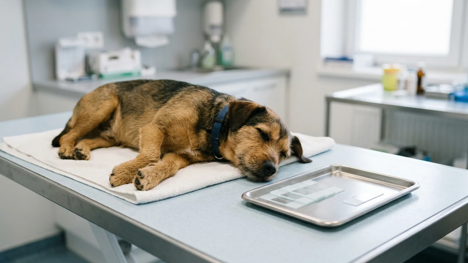

Urinalysis in Dogs and Cats: Reading Specific Gravity, Dipstick, and Sediment
How to read veterinary urinalysis results in dogs and cats: specific gravity cutoffs, valid dipstick pads, sediment findings, and what triggers next tests.

When your veterinarian orders a "complete urinalysis" (UA) for your dog or cat, they are not running a single test. A complete veterinary urinalysis is a three-part diagnostic assay evaluating the physical properties (color, clarity, and optical specific gravity), chemical constituents (via a dry reagent dipstick pad), and microscopic sediment (centrifuged cells, crystals, casts, and microbes) from a single urine specimen.
Along with the Complete Blood Count (CBC) and the Serum Blood Chemistry Panel, the urinalysis forms the third pillar of the veterinary Minimum Database. It is the single most cost-effective diagnostic tool in small-animal medicine for evaluating kidney function, localizing lower urinary tract disease, detecting systemic metabolic disorders, and monitoring therapeutic interventions.
However, interpreting a veterinary urinalysis report requires navigating two critical realities that pet owners are rarely told:
How the sample was collected completely changes what the numbers mean. A free-catch sample collected off the floor or grass carries background bacterial and cellular contamination that can mimic severe infection in a healthy pet.
Several standard human dipstick pads are medically invalid in dogs and cats. Pads for urine specific gravity, leukocyte esterase, nitrite, and urobilinogen produce notorious false positives and false negatives in small animals and must be ignored in favor of optical refractometry and microscopic sediment examination.
| Urinalysis Section | Target Analytes & Metrics | Primary Clinical Meaning | Key Small-Animal Nuance |
|---|---|---|---|
| Physical Exam | Color, Clarity, Urine Specific Gravity (USG) | Measures renal tubular concentrating ability and hydration status | Must use optical refractometer; dipstick USG pad is invalid. Human refractometers overestimate cat USG by 0.002–0.005. |
| Chemical Dipstick | pH, Protein, Glucose, Ketones, Bilirubin, Blood/Heme | Screens for metabolic spillover, proteinuria, hemolysis, and acid-base shifts | Leukocyte esterase and nitrite pads are invalid in dogs/cats. Bilirubin is normal in concentrated male dog urine; always abnormal in cats. |
| Microscopic Sediment | RBCs, WBCs, Epithelial cells, Crystals, Casts, Bacteria | Localizes inflammation, bleeding, nephron damage, and crystalluria | Analyzed on centrifuged sediment. >5 RBC/HPF or >5 WBC/HPF is abnormal. Crystals can form in vitro during refrigeration. |
What Is a Complete Urinalysis in Dogs and Cats?
A proper veterinary urinalysis must never be reduced to a quick dipstick check in an exam room. Urine is a complex aqueous solution containing metabolic waste products, electrolytes, exfoliated epithelial cells, and organic solutes. A full evaluation follows a standardized three-tier workflow:
        THE THREE-TIER VETERINARY URINALYSIS PROTOCOL

  ┌──────────────────────────────────────────────────────────────────┐
  │ TIER 1: PHYSICAL PROPERTIES & OPTICAL REFRACTOMETRY              │
  ├──────────────────────────────────────────────────────────────────┤
  │ • Visual inspection: Color (yellow, amber, red, brown) & Clarity │
  │ • Optical Refractometer USG: Measures total solute concentration │
  └─────────────────────────────────┬────────────────────────────────┘
                                    │
  ┌─────────────────────────────────▼────────────────────────────────┐
  │ TIER 2: CHEMICAL REAGENT DIPSTICK EVALUATION                     │
  ├──────────────────────────────────────────────────────────────────┤
  │ • Valid pads in pets: pH, Protein, Glucose, Ketones, Bilirubin,  │
  │   Occult Blood/Heme                                              │
  │ • Invalid pads to ignore: Leukocytes, Nitrites, Dipstick USG     │
  └─────────────────────────────────┬────────────────────────────────┘
                                    │ Centrifugation (1,000–1,500 rpm)
  ┌─────────────────────────────────▼────────────────────────────────┐
  │ TIER 3: MICROSCOPIC SEDIMENT EXAMINATION                         │
  ├──────────────────────────────────────────────────────────────────┤
  │ • Low Power Field (10x / LPF): Casts, squamous cells, crystals   │
  │ • High Power Field (40x / HPF): RBCs, WBCs, bacteria, lipid      │
  └──────────────────────────────────────────────────────────────────┘
Why the Collection Method Changes What the Results Mean
Before looking at a single number on a lab report, a veterinarian first checks the line labeled Collection Method. In small-animal practice, urine is obtained through three distinct techniques, each carrying unique strengths and diagnostic artifacts:
         COMPARISON OF VETERINARY URINE COLLECTION TECHNIQUES

  [ CYSTOCENTESIS ] (The Gold Standard)
  • Sterile needle aspiration directly through the abdominal wall into bladder.
  • Zero distal urethral, prepucial, or vaginal contamination.
  • REQUIRED for quantitative urine culture and susceptibility (C&S).
  • Artifact: Mild iatrogenic microscopic hematuria (a few extra RBCs)
    from needle puncture of the bladder wall is common.

  [ VOIDED / FREE-CATCH ] (Midstream Catch)
  • Collected in a clean cup or non-absorbent litter as the pet urinates.
  • Excellent for initial USG, glucose, and screening.
  • Artifact: Sweeps up squamous cells, bacteria, and lipid from the distal
    urethra, vulva, prepuce, and skin hair. Not reliable for sterile culture.

  [ CATHETERIZATION ] (Transurethral Catheter)
  • Flexible catheter passed through urethra into bladder.
  • Useful for relieving obstructions and monitoring hospitalized ICU output.
  • Artifact: Induces mechanical sloughing of transitional epithelial sheets
and RBCs. Induces bacterial colonization in 10%–55% of catheterized pets.| Collection Technique | Sterile for Bacterial Culture? | Distal Flora & Mucus Contamination? | Expected Mechanical Artifacts | Best Clinical Application |
|---|---|---|---|---|
| Cystocentesis | Yes (Reference Standard) | None | Mild iatrogenic microscopic hematuria (a few extra RBCs) | Suspected UTI, culture & sensitivity, definitive proteinuria workup |
| Free-Catch (Voided) | No | High (distal urethra, vulva, prepuce, hair) | Elevated squamous epithelial cells, background bacteria, lipid droplets | Baseline wellness screening, serial glucose/ketone tracking, USG |
| Catheterization | Conditional (if placed aseptically) | Moderate | Traumatic hematuria, large cohesive sheets of transitional epithelium | Urethral obstruction relief, ICU urine output monitoring |
The Sample Freshness Rule: Temperature, Time, and Artifacts
Urine is biologically active and unstable once outside the body. As urine sits, chemical and cellular alterations begin immediately:
At Room Temperature: Within 60 minutes, urea-splitting bacteria proliferate and consume urea, driving urine pH alkaline. Elevated pH causes red blood cells and white blood cells to lyse—research shows WBC counts decline by approximately 50% after one hour at room temperature (Yadav et al., 2020). Cellular casts dissolve, and glucose is metabolized by bacteria.
Under Refrigeration (2°C to 8°C): Refrigeration preserves cell morphology and inhibits bacterial multiplication for up to 6 hours. However, refrigeration increases urine specific gravity slightly and dramatically reduces solute solubility, causing in vitro crystallization of struvite and calcium oxalate dihydrate crystals that were not present in the living bladder.
Clinical Takeaway: Urine should ideally be analyzed within 30 to 60 minutes of collection. If delayed, it must be refrigerated immediately in a sealed, sterile container and warmed gently to room temperature before testing. Never freeze a urine sample destined for urinalysis or culture.
Reading Urine Specific Gravity: What 1.008–1.012 Means
Urine Specific Gravity (USG) is the ratio of the weight of a urine volume compared to an equal volume of pure distilled water (defined as 1.000). It measures the total concentration of dissolved solutes (primarily urea, creatinine, sodium, potassium, and chloride) and evaluates the functional capacity of renal tubules to concentrate or dilute glomerular filtrate.
USG must always be measured using a temperature-compensated optical refractometer. The dry reagent chemical strip pad for USG is formulated exclusively for human ionic strength and is notoriously inaccurate in domestic animals.
                  SPECIES USG INTERPRETATION BANDS

  1.000 ────────── 1.007 ────────── 1.012 ────────── 1.030 ────────── 1.085+
    │   HYPOSTHENURIA │  ISOSTHENURIA   │ MINIMALLY  │ ADEQUATE CONCENTRATION
    │   (Active Renal │  (Exact plasma  │ CONCENT-   │ (Healthy functional
    │    Dilution)    │   osmolality)   │ RATED      │  nephrons)
    ▼                 ▼                 ▼            ▼
  Diluted below     Tubules did no    Incomplete     • Dogs:  ≥ 1.030
  plasma (<1.007).  work (1.008–      concentrating  • Cats:  ≥ 1.035
  Seen in diabetes  1.012). Critical  filtration.   (Can exceed 1.080 in
  insipidus, psych- red flag if paired               concentrating cats)
  ogenic polydipsia with azotemia.
The Four USG Diagnostic Categories
Hyposthenuria (USG < 1.007): The urine is significantly more dilute than glomerular filtrate (plasma osmolality). This proves that the renal tubules are functional and actively expelling solute while retaining water. Causes include central or nephrogenic diabetes insipidus, psychogenic polydipsia, hyperadrenocorticism (Cushing's), hypercalcemia, and pyometra.
Isosthenuria (USG 1.008 to 1.012): The specific gravity of the urine is identical to protein-free plasma filtrate. The renal tubules have performed neither net concentration nor net dilution.
In a well-hydrated animal that just drank water: A single isosthenuric reading can be normal biological fluctuation.
In a dehydrated or azotemic animal: Isosthenuria is a hallmark of primary renal failure. If blood urea nitrogen (BUN) and creatinine are elevated on a blood chemistry panel while the urine is isosthenuric, it proves the kidneys have lost more than 66% to 75% of functional concentrating capacity.
Minimally Concentrated Urine (USG 1.013 to 1.029 in Dogs; 1.013 to 1.034 in Cats): The kidneys are concentrating urine above plasma level, but not reaching optimal physiological thresholds. Commonly seen in early chronic kidney disease, osmotic diuresis (diabetes mellitus), diuretic therapy, or liver dysfunction.
Adequate Renal Concentrating Ability (USG ≥ 1.030 in Dogs; ≥ 1.035 in Cats): Confirms that functional nephrons are actively resorbing water under the influence of antidiuretic hormone (ADH). Healthy cats often concentrate urine between 1.045 and 1.085, with a species ceiling of about 1.085.
The Feline Refractometry Scale Artifact
A crucial clinical pathology nuance highlighted by Yadav et al. (2020) is that feline urine contains higher concentrations of proteins and chromogens than canine or human urine. When cat urine is read on a standard human-calibrated refractometer, the refractive index overestimates feline USG by approximately 0.002 to 0.005. Modern veterinary diagnostic laboratories utilize veterinary-specific refractometers with dedicated feline optical scales to prevent misclassifying a cat with mild concentrating impairment as completely normal.
Which Dipstick Pads Matter in Pets — and Which Don't
A standard urinalysis dipstick contains 8 to 10 chemical reagent pads impregnated with colorimetric indicators. Because these strips were engineered for human medicine, several pads are clinically misleading in dogs and cats.
VETERINARY REAGENT DIPSTICK VALIDITY & INTERPRETATION
| VALID & ACTIONABLE IN PETS | INVALID / UNRELIABLE IN PETS |
|---|---|
| [✓] pH (5.5–7.5 baseline) | [X] Leukocyte Esterase Pad |
| [✓] Protein (Albumin-sensitive) | • False positives in cats |
| [✓] Glucose (Renal threshold) | • False negatives in dogs |
| [✓] Ketones (Acetoacetate) | [X] Nitrite Pad |
| [✓] Bilirubin (Species-specific) | • High false negative rate |
| [✓] Occult Blood / Heme | [X] Dipstick USG Pad |
| • Must use refractometer |
| Dipstick Pad | Diagnostic Validity in Small Animals | Mechanism & Reference Values | Common Causes of Abnormal Results |
|---|---|---|---|
| pH | Valid | Measures hydrogen ion activity. Normal carnivore urine is acidic (5.5 to 7.0). Post-prandial alkaline tide can reach 7.5. | Alkaline (>7.5): Infection with urease-producing bacteria (Staphylococcus, Proteus), vegetarian diets, stale stored sample. Acidic (<5.5): Metabolic acidosis, high-protein meat diet, catabolic states. |
| Protein | Valid (Interpret with USG) | Tetrabromophenol blue dye. Highly sensitive to albumin, insensitive to globulins or Bence-Jones proteins. | Prerenal: Hemoglobinuria, myoglobinuria, severe hyperglobulinemia. Renal: Glomerular disease, tubular dysfunction. Postrenal: Hematuria, pyuria, active cystitis. |
| Glucose | Valid | Glucose oxidase enzymatic reaction. Normal is negative. Reabsorbed in proximal tubules. | Hyperglycemic Glucosuria: Exceeds renal threshold (~200 mg/dL in dogs; 250–300 mg/dL in cats) in diabetes mellitus or severe stress. Euglycemic Glucosuria: Normal blood sugar; proximal tubulopathy (Fanconi syndrome, nephrotoxins). |
| Ketones | Valid | Nitroprusside reaction detecting acetoacetate and acetone (does not detect beta-hydroxybutyrate). | Diabetic ketoacidosis (DKA), prolonged starvation, severe hypoglycemia, high-fat ketogenic diets. |
| Bilirubin | Valid (Species Nuance) | Diazonium salt reaction. Detects conjugated bilirubin. | Dogs: Male dogs have low renal threshold and renal tubular conjugating capacity; trace to 1+ in concentrated urine (USG > 1.030) is normal. Cats: High renal threshold; ANY bilirubin in cat urine is pathological (hepatic lipidosis, hemolysis, cholangitis). |
| Occult Blood / Heme | Valid (Pseudoperoxidase) | Detects pseudoperoxidase activity of intact RBCs, free hemoglobin, and myoglobin. | Speckled pattern: Intact RBCs (true hematuria). Solid diffuse color: Free hemoglobin (hemolytic anemia) or myoglobin (rhabdomyolysis/muscle trauma). |
| Leukocyte Esterase | INVALID (Do Not Use) | Engineered for human neutrophil esterase enzymes. | Frequently false-positive in cats (even without pyuria) and false-negative in dogs (even when pyuria is present). Always use microscopic sediment examination to evaluate white blood cells. |
| Nitrite | INVALID (Do Not Use) | Requires dietary nitrate and specific nitrate-reducing bacteria over 4+ hours of bladder incubation. | High false-negative rate in carnivores due to low dietary nitrate and rapid voiding. |
Interpreting Protein in Context: The USG Multiplier
A dipstick result of "Trace" or "1+ Protein" cannot be evaluated in isolation:
In a cat with highly concentrated urine (USG 1.060), a trace or 1+ protein reading represents normal physiological background secretion (Tamm-Horsfall mucoprotein) concentrated in minimal water.
In an animal with dilute urine (USG 1.010), a 1+ protein reading indicates substantial, pathologically significant renal protein loss that warrants quantification with a Urine Protein-to-Creatinine (UPC) Ratio.
What RBCs, WBCs, Crystals, and Casts in the Sediment Mean
After physical and chemical analysis, 5 mL of urine is centrifuged at 1,000 to 1,500 rpm for 5 minutes. The supernatant is decanted, and the concentrated sediment pellet is examined under light microscopy at low power (10x objective / LPF) and high power (40x objective / HPF).
        SEDIMENT MICROSCOPY: NORMAL VS PATHOLOGICAL THRESHOLDS

  CELLS & ARTIFACTS (High Power Field - 40x / HPF):
  • Red Blood Cells (RBCs):  Normal: 0–5 / HPF   |  >5 / HPF: Hematuria
  • White Blood Cells (WBCs):Normal: 0–5 / HPF   |  >5 / HPF: Pyuria / Inflammation
  • Squamous Epithelial:     Normal: Few in voided samples (distal urethra)
  • Transitional Epithelial: Normal: Rare / HPF  |  Sheets: Catheter trauma / Neoplasia
  • Renal Tubular Epithelial:Normal: None        |  Present: Acute tubular injury

  CASTS (Low Power Field - 10x / LPF):
  • Hyaline Casts:           Normal: 0–2 / LPF   |  Proteinaceous tubular mold
  • Granular Casts:          Normal: None        |  Active degenerating tubular necrosis
  • Cellular / Waxy Casts:   Normal: None        |  Severe chronic intrarenal stasis

  CRYSTALS:
  • Struvite (Magnesium Ammonium Phosphate): Often incidental; alkaline pH artifact
  • Calcium Oxalate Dihydrate (Envelope):     Often incidental; acidic pH
  • Calcium Oxalate Monohydrate (Picket):     Ethylene glycol (antifreeze) toxicity
  • Ammonium Urate (Thorn-Apple):             Portosystemic shunt, Dalmatian defect
  • Cystine (Hexagonal Plates):               Genetic proximal tubule transporter defect
1. Cellular Constituents
Red Blood Cells (RBCs): Normal reference is 0 to 5 RBC/HPF. Elevated counts (>5/HPF) define hematuria, originating from hemorrhage, urolithiasis, neoplasia (transitional cell carcinoma), infection, or iatrogenic needle puncture during cystocentesis. If your dog has visible bloody urine, see our dedicated triage on dog blood in urine (hematuria).
White Blood Cells (WBCs): Normal reference is 0 to 5 WBC/HPF. Elevated counts (>5/HPF) define pyuria, indicating active inflammation or infection in the kidneys, ureters, bladder, urethra, or prostate.
Epithelial Cells:
Squamous cells: Large, polygonal cells with small nuclei from the distal urethra and genital tract. Expected in free-catch samples; absent in cystocentesis.
Transitional cells: Round to pear-shaped cells lining the renal pelvis, ureters, and bladder. Marked clusters with nuclear atypia raise concern for urothelial carcinoma.
Renal tubular epithelial cells: Small round cells from the proximal/distal tubules. Their presence indicates acute renal parenchymal insult.
2. Urinary Casts (Cylindruria)
Casts are cylindrical molds formed in the lumens of the distal convoluted tubules and collecting ducts, composed of a mucoprotein matrix (uromodulin / Tamm-Horsfall protein). Because they form exclusively inside nephrons, casts localize disease directly to the renal parenchyma:
Hyaline Casts: Pure protein molds. 0 to 2 per LPF can be normal after strenuous exercise or fever.
Granular Casts: Formed when cellular debris breaks down inside tubules. A marker of active acute tubular injury or nephrotoxicity (e.g., aminoglycoside or NSAID toxicity).
Cellular Casts (RBC or WBC Casts): Indicate active intrarenal hemorrhage (glomerulonephritis) or active renal infection (pyelonephritis).
Waxy Casts: Smooth, opaque casts with squared ends, representing the final stage of granular cast degeneration. They indicate prolonged tubular transit time and severe chronic intrarenal stasis.
3. Urinary Crystals (Crystalluria)
Crystalluria is the precipitation of mineral solutes in urine. Crucially, the presence of crystals does not equal a bladder stone (urolith), nor does it automatically require medical treatment:
                  CRYSTAL IDENTIFICATION GUIDE

  STRUVITE (Magnesium Ammonium Phosphate)
  • Appearance: Colorless, rectangular "coffin-lid" prisms.
  • Urine pH: Alkaline (>7.0).
  • Meaning: Very common in concentrated, refrigerated samples. In dogs,
    strongly associated with urease-producing bacterial UTIs. In cats,
    commonly sterile and diet-associated.

  CALCIUM OXALATE DIHYDRATE
  • Appearance: Colorless, square "envelope" with intersecting diagonal lines.
  • Urine pH: Acidic to neutral (<6.5).
  • Meaning: Can be normal in healthy pets. Increased numbers seen in hyper-
    calcemia, hyperoxaluria, or predisposing stone-forming conditions.

  CALCIUM OXALATE MONOHYDRATE
  • Appearance: "Picket fence" slats, dumbbell, or hemp-seed shapes.
  • Urine pH: Acidic.
  • Meaning: Pathognomonic marker for ETHYLENE GLYCOL (antifreeze) TOXICITY.

  AMMONIUM URATE / BIURATE
  • Appearance: Yellow-brown "thorn-apple" spheres with irregular spicules.
  • Urine pH: Acidic to neutral.
  • Meaning: Associated with congenital portosystemic vascular anomalies
    (liver shunts) and genetic transporter mutations in Dalmatians/Bulldogs.

  CYSTINE
  • Appearance: Perfect, colorless flat hexagonal plates (stop signs).
  • Urine pH: Acidic.
  • Meaning: Inborn genetic defect in renal tubular cystine reabsorption.
When Does a Urinalysis Change What the Vet Does Next?
A urinalysis is a clinical compass: it directs which diagnostic pathway to pursue and prevents inappropriate treatment.
       CLINICAL DECISION ROADMAP BASED ON URINALYSIS FINDINGS

  FINDING ON URINALYSIS                 NEXT CLINICAL ACTION
  ────────────────────────────────────────────────────────────────────────
  [ Azotemia + USG < 1.030/1.035 ]   →  Stage Chronic Kidney Disease (IRIS)
                                        Check blood pressure, SDMA, and UPC

  [ Glucosuria + Hyperglycemia ]     →  Verify Diabetes Mellitus (Fructosamine)
                                        Screen for ketoacidosis (urine ketones)

  [ Pyuria (>5 WBC) + Bacteria ]     →  Submit sterile Cystocentesis sample for
                                        Quantitative Culture & Susceptibility (C&S)

  [ Proteinuria on Dipstick (≥1+) ]  →  Exclude active sediment (RBC/WBC first)
                                        If sediment quiet, order quantitative UPC

  [ Hematuria + Negative Culture ]   →  Abdominal Ultrasound & Radiographs
                                        Evaluate for uroliths, TCC, FIC, trauma
The ISCAID Rule: Subclinical Bacteriuria vs. Active UTI
One of the most consequential modern consensus guidelines in veterinary medicine comes from the International Society for Companion Animal Infectious Diseases (ISCAID) (Weese et al., 2019):
"Subclinical bacteriuria is defined as the presence of bacteria in urine as determined by positive bacterial culture from a properly collected urine specimen, in the absence of clinical evidence of infectious urinary tract disease... Treatment of subclinical bacteriuria with antimicrobials is rarely indicated and is discouraged."
— ISCAID Guidelines, The Veterinary Journal (PMID: 30971357)
Finding bacteria on a sediment slide or a positive culture in a dog or cat that is urinating comfortably without straining, blood, or increased frequency does not constitute an infection requiring antibiotics. Treating subclinical bacteriuria does not prevent future infections and breeds multi-drug resistant bacterial strains. For clinical management of confirmed infections, see our guide to canine urinary tract infections and how to read culture and sensitivity reports.
Furthermore, a 2025 systematic review and meta-analysis by Thassakorn et al. (Animals, PMID 41375516) evaluating pooled data across dogs and cats with lower urinary tract signs demonstrated that bacterial infection accounts for only a subset of cases—reminding clinicians that sterile inflammatory conditions (such as feline idiopathic cystitis or uroliths) are frequent culprits.
How Should You Collect and Store a Urine Sample at Home?
If your veterinarian asks you to collect a free-catch sample from your dog or cat at home, proper handling prevents costly test errors:
        HOME URINE COLLECTION PROTOCOL FOR DOGS AND CATS

  FOR DOGS (Free-Catch Midstream):
  1. Use a clean, dry, non-metallic container (e.g., a clean soup ladle or
     shallow disposable plastic tray washed with hot water and no soap residue).
  2. Walk the dog on a short leash. When they squat or lift their leg, wait
     for the initial stream to pass (which clears distal urethral debris), then
     slide the collection container into the MIDSTREAM flow.
  3. Transfer 5–10 mL into a sterile, sealable container.

  FOR CATS (Non-Absorbent Litter):
  1. Empty and thoroughly wash a clean litter box with hot water (no bleach).
  2. Fill the box with non-absorbent hydrophobic plastic beads (e.g., Kit4Cat
     or Nosorb) or clean, dry aquarium gravel.
  3. Once the cat urinates, use the supplied pipette or clean syringe to aspirate
     the pooled urine from the bottom of the box into a clean tube.

  STORAGE AND TRANSPORT:
  • Keep the sample out of direct sunlight and heat.
  • Transport to the veterinary clinic within 30–60 minutes if possible.
  • If transport is delayed, store in the refrigerator (2°C–8°C) for up to 4–6
    hours maximum. Label the tube with your pet's name, date, and exact collection time.
Frequently Asked Questions
Can I collect my pet's urine sample at home?
Yes, for basic physical screening, specific gravity monitoring, or serial glucose/ketone checks. However, home-collected voided samples cannot be used for definitive sterile urine culture because they are contaminated by flora from the skin, hair, and distal urethra. If your veterinarian needs to confirm a bacterial infection or run an antibiotic sensitivity test, a sterile in-clinic cystocentesis sample is required.
Are crystals in a dog's urine an emergency?
No. Crystals (such as struvite or calcium oxalate dihydrate) frequently form ex vivo when urine cools down during transport or refrigeration. In an asymptomatic dog, crystalluria is often an incidental laboratory finding rather than an active disease. However, if your pet is straining, crying out, attempting to urinate without producing a stream, or passing blood, crystals may have consolidated into a bladder stone causing a life-threatening urethral blockage—which is an immediate medical emergency. For confirmed feline stones, see bladder stones in cats. For retail urinary-food claims, see urinary crystals and pet nutrition.
Why did my vet order a UPC ratio after the urinalysis?
The chemical dipstick pad only provides a semi-quantitative estimate of protein (Trace, 1+, 2+, 3+) and is biased toward albumin. Furthermore, dipstick protein must be evaluated against how concentrated the urine is. The Urine Protein-to-Creatinine (UPC) Ratio is a precise quantitative assay that divides total urinary protein by urinary creatinine, eliminating the confounding effect of urine dilution and providing an exact metric for glomerular kidney disease. Learn more in our UPC ratio and proteinuria guide.
What does a low urine specific gravity mean?
A low USG (<1.008 to 1.012) means the urine is dilute. If your pet has been drinking heavily after exercise, a transient low USG is normal. However, persistent low specific gravity indicates that the kidneys are unable to concentrate urine. Common causes include chronic kidney disease, diabetes insipidus, Cushing's disease, pyometra, hypercalcemia, liver failure, or psychogenic polydipsia. If your dog is drinking excessively, consult our guide on why your dog is drinking so much water.
Do dipstick 'blood' and 'leukocyte' results mean infection?
Not necessarily. The dipstick leukocyte esterase pad is completely invalid in dogs and cats and produces rampant false positives in cats. The dipstick blood pad detects chemical heme, reacting equally to intact red blood cells from mild bladder wall irritation, sterile stones, cancer, or microscopic needle trauma from collection. Infection is confirmed only when elevated white blood cells (pyuria) and bacteria are visibly confirmed under microscope sediment evaluation and verified by a sterile quantitative culture.
Sources
Yadav SN, Ahmed N, Nath AJ, Mahanta D, Kalita MK. Urinalysis in dog and cat: A review. Veterinary World. 2020;13(10):2133-2141. PMC Open Access: PMC7704312 | PubMed PMID: 33281347
Weese JS, Blondeau J, Boothe D, et al. International Society for Companion Animal Infectious Diseases (ISCAID) guidelines for the diagnosis and management of bacterial urinary tract infections in dogs and cats. The Veterinary Journal. 2019;247:8-25. PubMed PMID: 30971357 | ISCAID Guidelines Open PDF
Thassakorn P, et al. Prevalence of Bacterial Urinary Tract Infections in Dogs and Cats with Lower Urinary Tract Diseases and Other Illnesses: A Systematic Review and Meta-Analysis. Animals. 2025;15(23):3456. PubMed PMID: 41375516
Cornell University College of Veterinary Medicine. eClinPath: Urinalysis, Concentrating Ability, Chemical Constituents, Crystals, and Casts. Cornell eClinPath Reference
Merck Veterinary Manual. Urinalysis in Small Animal Clinical Pathology. Merck Veterinary Manual Urinalysis Chapter
Grauer GF. Proteinuria: measurement and interpretation. Topics in Companion Animal Medicine. 2011;26(3):121-127. PubMed PMID: 21782142